Skip to content
Dr. Khalid AletaibiConservative Dentistry · Dubai

BIOMIMETIC RESTORATIONS

Composite fillings, done the conservative way.

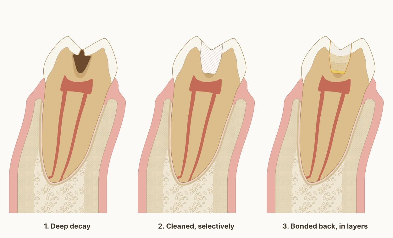
Three stages of a composite filling in a lower molar: deep decay with a dark infected core inside a lighter affected zone, then the cavity cleaned with a thin affected layer left over the nerve, then a glass-ionomer liner and composite bonded back in small layers.

A bonded composite filling, properly placed, is one of the most conservative restorations modern dentistry can offer. The difference between a routine filling and a long-lasting one is almost entirely a matter of how it is done.

Dr. Khalid AletaibiWritten by Dr. Khalid Aletaibi, DHA-licensed dentist

QUICK ANSWER

A composite filling repairs a cavity using a tooth-coloured resin that is bonded directly to the tooth. That bond is what makes it minimally invasive: only the decay is removed, where older materials had to be held in place by retentive shapes cut into sound tooth around them. When placed under careful isolation with modern adhesive technique, posterior composites have an annual failure rate of roughly 1-3%, with median survival comparable to amalgam in well-controlled studies. The conservative principle is simple: remove only what is diseased, preserve everything that is sound, and bond what remains.

Visits

Usually 1

Anaesthetic

Local, if needed

Tooth removed

Diseased tissue only

Reversibility

Repairable, replaceable

Longevity

~1-3% fail per year

Why I default to composite, the white filling

Composite is bonded to enamel and dentine. That single property changes everything: I can remove only the carious tissue and leave the sound structure alone, instead of cutting an undercut shape into the tooth to mechanically retain a non-bonded material. Less drilling means more remaining tooth. More remaining tooth means a stronger restored unit and a longer working life for the tooth itself, which is the only metric that ultimately matters.

Modern systematic reviews consistently report annual failure rates for posterior composites in the range of 1-3% [1][2]. A 2023 meta-analysis of randomised trials found that composite fillings built directly in back teeth may last as long as composite restorations made in a laboratory [3]. The number that matters in practice is not the average but the slope: failure rates climb when isolation is compromised, when caries is over- or under-removed, or when the restoration is built up in a single bulk increment without attention to polymerisation shrinkage.

The same lower first molar in every panel, cut from the cheek side to the tongue side through its front root, with the gum, ligament and bone around it. The tooth and bone are drawn to published measurements; the decay and the filling show what typically happens, not one patient's tooth.

Diagram page and reuse terms: A composite filling: only the decay comes out, and the tooth is bonded back in layers

Sources and measurements
  • Decay is called deep when it reaches the inner quarter of the dentine but still leaves a protective layer of hard or firm dentine over the pulp: the European Society of Endodontology's definition, restated in the 2026 S3-level guideline on deep caries from European dental societies. The guideline supports selective or stepwise removal over complete removal, to lower the risk of exposing the pulp, and with selective removal the dentine at the edges is still cleaned to hard, for a secure restoration. Schwendicke et al. 2026, Clinical Oral Investigations. DOI 1, A composite filling: only the decay comes out, and the tooth is bonded back in layers
  • The decay is drawn as two zones, the two layers of carious dentine often called infected and affected dentine. In the outer layer the cross-links that hold the dentine's collagen together were irreversibly destroyed; in the inner layer the change was considered reversible. Kuboki, Ohgushi and Fusayama 1977, Journal of Dental Research, laboratory study. How far each zone reaches in the drawing is illustrative, not measured. DOI 2, A composite filling: only the decay comes out, and the tooth is bonded back in layers
  • The decay's shape follows the textbook drawing of decay in a groove: small at the surface, widening toward the junction with the dentine, then wider in the dentine and on toward the pulp. Woelfel's Dental Anatomy, Figure 10-1.
  • In deep lesions in teeth with healthy pulps, international consensus recommends selective removal to soft dentine: decayed dentine close to the pulp does not need to be removed, and the priorities are keeping healthy tooth tissue, achieving a seal and keeping the pulp healthy. Schwendicke et al. 2016, Advances in Dental Research, International Caries Consensus Collaboration. DOI 4, A composite filling: only the decay comes out, and the tooth is bonded back in layers
  • A meta-analysis of 10 randomised trials with 1,257 patients found that leaving some decayed dentine over the pulp, in one visit or two, cut the odds of exposing the pulp to about a third of those with complete removal (odds ratio 0.31). Schwendicke, Dörfer and Paris 2013, Journal of Dental Research, systematic review and meta-analysis. DOI 5, A composite filling: only the decay comes out, and the tooth is bonded back in layers
  • In 32 teeth with deep decay, the decay was only partly removed, then the teeth were lined with calcium hydroxide and sealed for six to seven months. When they were reopened the dentine was dry, it was hard in 80 per cent of the 30 teeth still in the study, and the bacteria in it had decreased significantly. Maltz et al. 2002, Quintessence International. PubMed 6, A composite filling: only the decay comes out, and the tooth is bonded back in layers
  • The liner is glass ionomer, whose acid bonds chemically to the calcium of the tooth's mineral: in a laboratory study, about two-thirds of its acid groups were found bonded to hydroxyapatite. Yoshida et al. 2000, Journal of Dental Research. DOI 7, A composite filling: only the decay comes out, and the tooth is bonded back in layers
  • Glass ionomers release fluoride and can take it up again, acting as a fluoride reservoir. Wiegand, Buchalla and Attin 2007, Dental Materials, review. DOI 8, A composite filling: only the decay comes out, and the tooth is bonded back in layers
  • Acid on the enamel only, before a universal adhesive: in a randomised trial of 295 back-tooth fillings in 70 patients, fillings bonded this way showed significantly fewer defects at their edges after 24 months than those bonded with the adhesive alone. Barceleiro et al. 2023, Journal of Applied Oral Science. DOI 9, A composite filling: only the decay comes out, and the tooth is bonded back in layers
  • The clinical steps are drawn as Dr. Khalid works: under rubber dam; all the infected dentine removed, with the dentine at the edges cleaned to hard and the enamel to sound so that the filling seals; a layer of affected dentine left over the pulp under a glass-ionomer liner; acid on the enamel only before the adhesive; and the composite built in small layers, opaque where the dentine was and translucent where the enamel was. The layer left over the pulp, about 0.7 mm at its deepest, and the 0.4 mm liner are representative thicknesses, not measurements.
  • The enamel follows one measured dataset for this tooth, from cone-beam CT of 45 lower first molars: 1.58 mm at the front cheek-side cusp tip and 0.98 mm at the central fossa, its thinnest point. Zhang et al. 2026, BMC Oral Health. DOI 11, A composite filling: only the decay comes out, and the tooth is bonded back in layers
  • Two canals in the front root, as drawn, are found in 94.4 per cent of lower first molars, and three in 2.3 per cent, from a systematic review of 41 studies covering 18,781 teeth. de Pablo et al. 2010, Journal of Endodontics. DOI 12, A composite filling: only the decay comes out, and the tooth is bonded back in layers
  • Bone level: on cone-beam CT of 100 lower molar sites in 25 adults, the crest of the bone sat 2.03 to 2.11 mm below the enamel edge of first molars, with 1.14 to 1.19 mm of bone over the cheek side of the root near its neck. Othman 2026, The Saudi Dental Journal. DOI 13, A composite filling: only the decay comes out, and the tooth is bonded back in layers
  • The cheek-side cortical plate near the crest is drawn from cone-beam CT of lower first and second molars, where it measured 1.1 to 1.4 mm. Sayed et al. 2021, The Saudi Dental Journal. DOI 14, A composite filling: only the decay comes out, and the tooth is bonded back in layers
  • Further from the crest, on cone-beam CT of 100 people, between the lower second premolar and first molar: the cheek-side plate measured 1.93 mm at 4 mm below the enamel edge and 2.36 mm at 6 mm, and the tongue-side plate 2.18 and 2.09 mm. Gupta et al. 2025, BMC Oral Health. DOI 15, A composite filling: only the decay comes out, and the tooth is bonded back in layers
  • The gum is drawn 1.2 mm thick, within the 0.8 to 1.5 mm measured by ultrasound across lower teeth in 200 periodontally healthy men. Eger, Müller and Heinecke 1996, Journal of Clinical Periodontology. DOI 16, A composite filling: only the decay comes out, and the tooth is bonded back in layers
  • The tooth follows the textbook descriptions of a lower first molar rather than a symmetrical outline: the crown leans toward the tongue on its root, the front root's tip lies under the front cheek-side cusp, the roof of the pulp chamber sits at the level of the enamel edge with the floor in the root trunk, and the ligament around the root is drawn 0.2 mm wide, under the quarter of a millimetre the textbooks give, inside a thin lining of compact bone. Wheeler's Dental Anatomy, Physiology and Occlusion, 11th edition, and Woelfel's Dental Anatomy.
  • The roof of the pulp chamber sits at the level of the enamel edge, where it lay in 97 per cent of 100 lower molars measured on radiographs, and the drawing's distance from the cheek-side cusp tip to that roof, 6.36 mm, is the study's mean. Deutsch and Musikant 2004, Journal of Endodontics. DOI 18, A composite filling: only the decay comes out, and the tooth is bonded back in layers

Selective caries removal, and why it matters

For decades, dental schools taught that all softened, discoloured dentine had to be excavated until the cavity floor was hard. International consensus changed that. The 2016 ICCC consensus paper [4], the 2015 network meta-analysis preceding it [5], and the 2021 Cochrane review of cavitated lesion management [6] all converge on the same conclusion: in deep cavities close to the pulp, leaving a thin layer of softer affected dentine and sealing it under a well-bonded restoration produces fewer pulp exposures and better long-term tooth survival than aggressive complete excavation.

In plain language: digging deeper is not safer, and a healthy pulp is worth more than a textbook-clean cavity floor. This single change in technique has prevented an enormous number of unnecessary root canals worldwide. It is also one of the clearest examples of evidence updating practice, and of older training that should be retired.

A close-up from the same measured molar as the step-by-step figure, with a 1 mm scale. All the infected dentine is gone; a thin layer of affected dentine stays over the pulp, sealed under the liner and the composite, with firm dentine still between it and the pulp. Leaving it, rather than digging on, lowers the risk of exposing the pulp, and dentine sealed in this way has been seen to harden.

Diagram page and reuse terms: What is left over the nerve

Sources and measurements
  • In deep lesions in teeth with healthy pulps, international consensus recommends selective removal to soft dentine: decayed dentine close to the pulp does not need to be removed, and the priorities are keeping healthy tooth tissue, achieving a seal and keeping the pulp healthy. Schwendicke et al. 2016, Advances in Dental Research, International Caries Consensus Collaboration. DOI 1, What is left over the nerve
  • Decay is called deep when it reaches the inner quarter of the dentine but still leaves a protective layer of hard or firm dentine over the pulp: the European Society of Endodontology's definition, restated in the 2026 S3-level guideline on deep caries from European dental societies. The guideline supports selective or stepwise removal over complete removal, to lower the risk of exposing the pulp, and with selective removal the dentine at the edges is still cleaned to hard, for a secure restoration. Schwendicke et al. 2026, Clinical Oral Investigations. DOI 2, What is left over the nerve
  • A meta-analysis of 10 randomised trials with 1,257 patients found that leaving some decayed dentine over the pulp, in one visit or two, cut the odds of exposing the pulp to about a third of those with complete removal (odds ratio 0.31). Schwendicke, Dörfer and Paris 2013, Journal of Dental Research, systematic review and meta-analysis. DOI 3, What is left over the nerve
  • In 32 teeth with deep decay, the decay was only partly removed, then the teeth were lined with calcium hydroxide and sealed for six to seven months. When they were reopened the dentine was dry, it was hard in 80 per cent of the 30 teeth still in the study, and the bacteria in it had decreased significantly. Maltz et al. 2002, Quintessence International. PubMed 4, What is left over the nerve
  • The decay is drawn as two zones, the two layers of carious dentine often called infected and affected dentine. In the outer layer the cross-links that hold the dentine's collagen together were irreversibly destroyed; in the inner layer the change was considered reversible. Kuboki, Ohgushi and Fusayama 1977, Journal of Dental Research, laboratory study. How far each zone reaches in the drawing is illustrative, not measured. DOI 5, What is left over the nerve
  • The liner is glass ionomer, whose acid bonds chemically to the calcium of the tooth's mineral: in a laboratory study, about two-thirds of its acid groups were found bonded to hydroxyapatite. Yoshida et al. 2000, Journal of Dental Research. DOI 6, What is left over the nerve
  • Glass ionomers release fluoride and can take it up again, acting as a fluoride reservoir. Wiegand, Buchalla and Attin 2007, Dental Materials, review. DOI 7, What is left over the nerve
  • Drawn as Dr. Khalid works: the affected layer is sealed under a glass-ionomer liner, then the composite. The layer left, about 0.7 mm at its deepest, and the 0.4 mm liner are representative thicknesses, not measurements; the firm dentine and the pulp are read off the same measured tooth.

What I do differently

Every composite I place is done under rubber dam isolation. The dam is not a stylistic choice, it is the single most important variable for adhesive success. Saliva and humidity contamination, even briefly, will compromise the bond. With the field properly isolated, I remove decay selectively, etch only the enamel and then apply the adhesive in a controlled sequence, and build the composite in small layers to manage shrinkage stress. Anatomy is shaped before light-curing, then refined with finishing burs and discs.

It takes longer than a 'just put a filling in' visit. That extra time is the difference between a restoration that lasts a decade and one that fails in three years. None of this is exotic technique, it is mainstream contemporary cariology, applied carefully every single time.

A close-up of the filling's cheek-side edge, from the same measured molar as the step-by-step figure, with a 1 mm scale. Acid goes on only where the wall is enamel, the adhesive then covers enamel and dentine alike, and the composite goes in in small layers that copy the tooth: opaque where the dentine was, translucent where the enamel was.

Diagram page and reuse terms: The bonded edge

Sources and measurements
  • Acid on the enamel only, before a universal adhesive: in a randomised trial of 295 back-tooth fillings in 70 patients, fillings bonded this way showed significantly fewer defects at their edges after 24 months than those bonded with the adhesive alone. Barceleiro et al. 2023, Journal of Applied Oral Science. DOI 1, The bonded edge
  • In fillings at the necks of teeth, a meta-analysis of 10 randomised trials found less staining and fewer adaptation defects at the edges when the enamel was etched selectively before a self-etch adhesive, and fewer fillings lost at three years. Szesz et al. 2016, Journal of Dentistry, systematic review and meta-analysis. DOI 2, The bonded edge
  • Drawn as Dr. Khalid bonds: acid on the enamel only, then the adhesive over enamel and dentine, and the composite in small layers. Where the cavity wall meets the junction of enamel and dentine, the etched band stops and the opaque layers begin. The etched band and the adhesive are drawn wider than life so they can be seen; their widths are not measurements.
  • The enamel follows one measured dataset for this tooth, from cone-beam CT of 45 lower first molars: 1.58 mm at the front cheek-side cusp tip and 0.98 mm at the central fossa, its thinnest point. Zhang et al. 2026, BMC Oral Health. DOI 4, The bonded edge

Composite filling vs silver amalgam

Composite filling vs silver amalgam
Bonded compositeSilver amalgam
AppearanceTooth-coloured and blends with your natural enamel.Dark silver and visible when you talk or laugh.
Healthy tooth removedOnly the decay; the filling bonds to what remains.Often more, because the cavity is shaped to hold the filling mechanically.
How it stays in placeBonded to the tooth, which helps support the remaining structure.Held by the shape of the cavity, not bonded to the tooth.
Mercury contentNone.Contains mercury bound within a stable metal alloy.
RepairabilityCan often be repaired or added to.Usually removed and replaced entirely.
Where it fits todayThe usual choice for most modern fillings.Long-serving, but now rarely placed for new fillings.

What happens, step by step

  1. 1

    We isolate the tooth

    A rubber dam keeps the tooth dry and free of saliva, which is the single most important factor for a lasting bond.

  2. 2

    We remove only the decay

    We take out the diseased tissue selectively and leave sound tooth structure untouched, staying away from the pulp.

  3. 3

    We bond and layer

    We etch only the enamel and apply the adhesive, then build the composite in small layers to manage shrinkage stress.

  4. 4

    We shape and polish

    We sculpt the anatomy, check the bite, then finish and polish so the filling blends into the tooth.

Conservative profile

  • How invasiveLow
  • Healthy tooth preservedHigh
  • Pulp protected in deep cavitiesHigh
  • Permanence of tooth removalLow

COMMON QUESTIONS

What patients ask most.

How long does a composite filling last?
In well-controlled clinical studies, posterior composite restorations have annual failure rates of roughly 1-3%, which translates to a median survival of more than 10 years for many patients. The longevity depends heavily on isolation, adhesive technique, occlusion, and the patient's caries risk, not just on the brand of composite. In a pooled analysis of 2,816 composite fillings in back teeth, the main reasons for failure were new decay and fracture, and fillings in people at high decay risk failed more often [1]. That is where routine care comes in: at your check-up and cleaning, set at 4, 6 or 12 months for you, the edges of each filling are checked and cleaned, so new decay is found while it is still small. What happens at a cleaning is set out in your cleaning, step by step.
Are composite fillings as good as silver amalgam?
For most posterior cavities, yes. Modern composites placed with adhesive isolation perform comparably to amalgam in long-term studies, while preserving more tooth structure because they don't require an undercut preparation. The conservative trade-off favours composite in nearly every clinical scenario where it can be properly placed.
Will my filling look obvious?
A correctly shade-matched and polished composite is essentially invisible. The art of layering, using a slightly translucent enamel layer over an opaque dentine core, is what produces a restoration that disappears into the tooth. This is editorial work, not just technical work.
I've been told I need a crown. Could a composite work instead?
Sometimes, yes, and it's worth asking. For teeth that have lost moderate amounts of structure, a well-designed bonded composite or an indirect inlay/onlay can restore function while preserving more of the natural tooth than a full crown. Crowns remain the right answer for severely broken-down teeth, but they are not the default. A second opinion is one of the most conservative things you can do before agreeing to a crown.
Does the deep filling mean I'll need a root canal?
Not usually. The current evidence supports leaving a thin layer of softer affected dentine over the pulp in deep cavities, sealed under a bonded restoration, rather than digging until the pulp is exposed. This approach significantly reduces unnecessary root canal treatment and preserves pulp vitality. Whether your tooth needs root canal therapy depends on whether the pulp is already inflamed or infected, not on the cavity depth alone.
Can old amalgam fillings be replaced with composite?
Yes, but only when there is a reason. Replacing intact, sound amalgams purely for aesthetic reasons sacrifices healthy tooth structure and is rarely the conservative choice. When an old amalgam has decay around it, is fractured, or is leaking, replacement with a bonded composite is a clear indication and the right call.

Speak to us

Would you prefer we call you?

Leave your name and number and Dr. Khalid's team will call you back to answer your questions and find a time that suits you.

Need a filling, or a second opinion on one?

Whether it's a small new cavity or a large old restoration that needs review, we'll start with a careful examination and the most conservative answer the evidence supports.

Book a Consultation

← Biomimetic Restorations & Endodontics

Call Dr. Khalid directlyDiscuss your concerns and questions