Skip to content
Dr. Khalid AletaibiConservative Dentistry · Dubai

PREVENTIVE DENTISTRY

Gum disease treatment and deep cleaning, step by step.

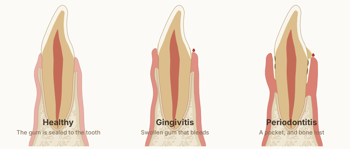
The same lower front tooth three times, cut from the lip side to the tongue side: healthy gum sealed to the tooth; gingivitis, a swollen gum that bleeds with the bone untouched; and periodontitis, a pocket with plaque and tartar on the root and bone lost.

Gum disease often gives few warning signs early on, so it is found by measuring rather than by waiting for something to hurt, and together with decay it is the main cause of tooth loss [1]. Dr. Khalid charts your gums, explains in plain words what stage they are at, and treats gingivitis and the earlier, shallower stages himself, starting with a careful deep cleaning where one is needed. Pockets deeper than 5 mm are treated by our periodontist in the same clinic, on one plan that Dr. Khalid, our periodontist and the team make together, so the finished result, agreed first, meets the standards of function, aesthetics and biomimetics; Dr. Khalid stays on your case throughout. The aim is the one the whole practice works to: keep your natural teeth, rebuild lost support where the evidence allows, and keep the disease from coming back.

Dr. Khalid AletaibiWritten by Dr. Khalid Aletaibi, DHA-licensed dentist

QUICK ANSWER

Gum disease treatment starts with a full chart: pocket depths at six points around every tooth, bleeding, and X-rays of the bone, so the disease can be given a stage and a grade [2]. The core treatment is a deep cleaning, known clinically as scaling and root planing: tartar and bacterial film are cleaned off the root surfaces below the gum under local anaesthetic. In the review behind the European guideline, deep cleaning made pockets about 1.4 mm shallower on average and closed about three in four of them at 6 to 8 months [3]. Every site is then re-measured, sites that stay deep go on to a further step, and supportive care every 3 to at most 12 months keeps the result [4].

Anaesthetic

Local, for deep cleaning

Deep cleaning visits

One to four, by approach

First treatment

Non-surgical

Pockets deeper than 5 mm

Our periodontist, on one joint plan

Supportive care

Every 3 to 12 months

What periodontitis is, in plain words

Periodontitis, still sometimes called pyorrhoea, is a long-running inflammation of the tissues that hold each tooth in its socket. It starts with the bacterial film called plaque at the gum line. In many mouths that film causes gingivitis, gums that are red and bleed easily, and goes no further. In a susceptible person the inflammation reaches deeper, and the fibres and bone that anchor the tooth are slowly lost. The gap this opens between the gum and the root is the pocket that the probe measures.

Susceptibility is partly inherited: the genetic share of the risk is estimated at up to half. The rest is acquired, and much of it can be changed: smoking, diabetes that is undiagnosed or not well controlled, and obesity all add to it [1]. That is why two people with the same amount of plaque can have very different gums, and why your chart, not the look of your gums, decides what you need.

Severe periodontitis is common. It affects about 11% of adults worldwide, and new cases are most frequent at around the age of 38 [5]. Your thirties are a sensible time to have your gums properly charted, even if nothing hurts and your teeth feel firm.

The reading is the distance from the gum's edge to where the probe stops, shown by the bracket beside each probe. Healthy gums read 1 to 3 mm and do not bleed; the aim of treatment is no pocket deeper than 4 mm that bleeds and none of 6 mm or more.

Diagram page and reuse terms: What the probe measures

Sources and measurements
  • Healthy gum anatomy: the gum edge sits about 1 mm above the enamel's edge and healthy gums probe 1 to 3 mm (Woelfel's Dental Anatomy). Under a groove about 0.7 mm deep lie about 1 mm of attaching epithelium and 1 mm of fibres above the bone, Gargiulo's autopsy means, which vary from person to person. DOI 1, What the probe measures
  • Gum thickness: at the lower front teeth the gum averaged 0.71 mm thick, and healthy pockets read 1.79 mm on average, in 187 people with healthy gums. DOI 2, What the probe measures
  • The probe: commercial probe tips measured 0.28 to 0.70 mm across, and a light force of about 0.25 N is used; at higher forces healthy gums bleed more often. DOI 3, What the probe measures
  • Where the probe stops in health: at 22 sites on front teeth, the probe tip stopped inside the junctional epithelium, on average 0.25 mm short of its end. DOI 4, What the probe measures
  • Where the probe stops in a pocket: in untreated pockets the tip went 0.29 mm (Magnusson and Listgarten 1980) and 0.45 mm (Fowler 1982) past the epithelium into inflamed tissue, well short of the bone; after treatment it stopped inside the epithelium. DOI 5, What the probe measures
  • Bone around lower central incisors: in CBCT scans of 50 women without gum disease, the bone crest lay about 2.2 to 2.8 mm below the enamel's edge, and the bone on the lip side was thin, about 0.5 to 0.75 mm including the ligament, along most of the root. DOI 6, What the probe measures
  • The European guideline (EFP S3): the aim is no pocket deeper than 4 mm that bleeds and none of 6 mm or more; sites are re-assessed after healing; hand or powered instruments, or both; supportive care every 3 to at most 12 months. DOI 7, What the probe measures
  • The tooth: a lower central incisor in section, crown 9.5 mm and root 12.5 mm (Wheeler's Dental Anatomy, Table 7.1), with the lip side on the left. Pockets and openings are drawn slightly open so they can be seen; no number on the drawing is a measurement of one person.

Stage and grade: the two labels on your chart

Since the 2017 world classification, periodontitis is described with two labels rather than one vague diagnosis [6]. The stage, from I to IV, records how much support has already been lost and how complex the case is to treat: how far the attachment loss reaches, how much bone the X-ray shows as lost, whether teeth have already been lost to the disease, and features such as deep pockets, vertical bone defects or loose teeth [2]. The table further down this page sets the four stages side by side.

The grade, A, B or C, describes speed: how fast the disease appears to be moving, judged from earlier X-rays where they exist and from the pattern of loss where they do not [2]. Grade B, moderate progression, is the starting point until the evidence moves it. Two risk factors push a case towards grade C, the fastest: smoking 10 or more cigarettes a day, and diabetes with an HbA1c of 7.0% or more [2][4].

Put simply, the stage tells you how much has been lost and the grade how quickly it is being lost. Both are written into your notes, so the next examination has something firm to be compared against, and you can see for yourself whether treatment is working.

Deep cleaning, step by step

Deep cleaning is the first active treatment for periodontitis. Its clinical names are scaling and root planing, or subgingival instrumentation. It is a different treatment from a routine scale and polish: a routine clean removes plaque, tartar and stain above the gum and just at its edge, while a deep cleaning reaches down the root surfaces inside the pockets, where the bacteria that drive the disease live.

First, the area is numbed with local anaesthetic, so the cleaning below the gum can be done thoroughly and comfortably. The root surfaces are then cleaned with an ultrasonic scaler, a fine tip that vibrates in a spray of water, and with hand instruments where a finer touch is needed. The European guideline's review found no difference between hand and powered instruments, and the guideline supports either or both [3][4]. The mouth can be treated a quarter at a time over several visits, or all at once within 24 hours; both gave similar results, so the choice is made around your mouth, your comfort and your diary [3][4].

What it achieves: in that review, pockets were on average about 1.4 mm shallower and about three in four had closed at 6 to 8 months [3], and an American Dental Association review of 72 studies found an average attachment gain of about 0.5 mm [7]. Antibiotic tablets are not a routine part of it: the guideline advises against using them routinely [4]. Dr. Khalid carries out the deep cleaning for the earlier, shallower stages himself. Sites with pockets deeper than 5 mm are treated by our periodontist in the same clinic, on the same plan, which Dr. Khalid, our periodontist and the team make together from your chart so the finished result, agreed first, meets the standards of function, aesthetics and biomimetics; Dr. Khalid stays on your case.

A close-up of the tongue side of the 5 mm pocket, with a 1 mm scale. Plaque and tartar stop short of the bone: below them lie a short seal of gum, a band of lost fibres, then fibres still holding the tooth.

Diagram page and reuse terms: Inside a pocket

Sources and measurements
  • Plaque never reaches the bone: on 48 extracted teeth the front of the plaque lay 0.2 to 2.0 mm from the remaining attachment fibres, and in autopsy material never less than 0.5 mm or more than 2.7 mm from the bone. DOI 1, Inside a pocket
  • Inside a pocket, between the last epithelium and the first intact fibres there was a zone of destroyed fibres averaging 0.43 mm wide, in almost every tooth examined. DOI 2, Inside a pocket
  • The pocket lining does not attach to the tooth: it is thin, ridged and full of inflammatory cells, with breaks in places (a study of experimental pockets in dogs). DOI 3, Inside a pocket
  • Tartar always carries plaque: a living film of plaque covers mineralised tartar deposits, above and below the gum. DOI 4, Inside a pocket
  • Two kinds of tartar: above the gum it is yellow-white, layered and about 37% mineral by volume; below the gum it is dark brown, uniform and about 58% mineral, usually a thin, smooth veneer on the root (Friskopp and Isacsson 1984; Woelfel; Gurgan and Bilgin 2005). DOI 5, Inside a pocket
  • Where the probe stops in a pocket: in untreated pockets the tip went 0.29 mm (Magnusson and Listgarten 1980) and 0.45 mm (Fowler 1982) past the epithelium into inflamed tissue, well short of the bone; after treatment it stopped inside the epithelium. DOI 6, Inside a pocket
  • The tooth: a lower central incisor in section, crown 9.5 mm and root 12.5 mm (Wheeler's Dental Anatomy, Table 7.1), with the lip side on the left. Pockets and openings are drawn slightly open so they can be seen; no number on the drawing is a measurement of one person.

The four steps of treatment

The European Federation of Periodontology's guideline sets treatment out in four steps, and each step starts only once the one before it has been checked [4]. Step 1 is for everyone: coaching on cleaning at home, with interdental brushes where the gums are inflamed, help with risk factors such as smoking and blood sugar, and a professional clean above the gum line. Gingivitis is treated with this kind of care, and gum health can be restored [8]. Step 2 is the deep cleaning described above.

After the deep cleaning, the gums are given time to heal, and every site is measured again about three months later. The target is clear: no pocket deeper than 4 mm that bleeds, and none of 6 mm or more [4]. Sites that reach it go straight on to supportive care. Sites that do not go on to step 3: a repeat deep cleaning, or gum surgery, in which the gum is gently opened so the root can be cleaned under direct vision and the pocket reduced. Where bone has been lost in a vertical, crater-like defect 3 mm or deeper, the guideline recommends regenerative surgery, which aims to rebuild part of the lost support [4].

In step 3, pockets deeper than 5 mm and any surgery are treated by our periodontist in the same clinic, and the work is planned before anything is booked: Dr. Khalid, our periodontist and the team plan the case together from the same chart and X-rays, so the finished result meets the standards of function, aesthetics and biomimetics. That result is agreed first, so the surgery is planned to the finished tooth rather than the tooth fitted to whatever the surgery leaves, and our periodontist carries out the surgery. Dr. Khalid stays on your case throughout, does the restorative work, and they review the result together. Step 4, supportive care, then keeps the result, with visits every 3 to at most 12 months [4].

The pocket closes mostly by a long seal of gum along the cleaned root, plus a little shrinkage of the gum edge; bone already lost does not grow back by itself. On average pockets were 1.4 mm shallower and about three in four closed at 6 to 8 months.

Diagram page and reuse terms: Deep cleaning and healing, step by step

Sources and measurements
  • What deep cleaning achieves: in the systematic review behind the European guideline, pockets were on average 1.4 mm shallower and about three in four had closed at 6 to 8 months. DOI 1, Deep cleaning and healing, step by step
  • How a pocket heals after root planing: by a long junctional epithelium along the cleaned root, with no new connective tissue attachment and no new bone (monkey studies; in humans, five of six control teeth showed the same at three months, Yukna 2007). DOI 2, Deep cleaning and healing, step by step
  • How much of the change is gum shrinkage: in 68 patients, 4 to 6 mm sites became 1.03 mm shallower with a 0.69 mm gain in measured attachment, so about a third of the change was the gum edge settling lower. DOI 3, Deep cleaning and healing, step by step
  • Where the probe stops in a pocket: in untreated pockets the tip went 0.29 mm (Magnusson and Listgarten 1980) and 0.45 mm (Fowler 1982) past the epithelium into inflamed tissue, well short of the bone; after treatment it stopped inside the epithelium. DOI 4, Deep cleaning and healing, step by step
  • The European guideline (EFP S3): the aim is no pocket deeper than 4 mm that bleeds and none of 6 mm or more; sites are re-assessed after healing; hand or powered instruments, or both; supportive care every 3 to at most 12 months. DOI 5, Deep cleaning and healing, step by step
  • Using an ultrasonic tip: the side of the last 1 to 2 mm is held close to parallel to the tooth with light pressure (maker's instructions; root damage was least at angles close to 0 degrees, Flemmig 1998). The tip moves only about 5 to 44 micrometres (Lea 2003). DOI 6, Deep cleaning and healing, step by step
  • The tooth: a lower central incisor in section, crown 9.5 mm and root 12.5 mm (Wheeler's Dental Anatomy, Table 7.1), with the lip side on the left. Pockets and openings are drawn slightly open so they can be seen; no number on the drawing is a measurement of one person.

Who does what: one plan, in the same clinic

Dr. Khalid examines and charts your gums, gives you the stage and grade, and treats gingivitis and the earlier, shallower stages of periodontitis without surgery: the coaching, the professional cleaning and the deep cleaning. He also provides your supportive care visits, and he carries out every cleaning himself.

Pockets deeper than 5 mm, gum surgery, regeneration and gum grafts are treated by our periodontist, who works in the same clinic. Whatever the stage, there is one plan: Dr. Khalid, our periodontist and the team plan your case together before anything is booked, from the same charts, X-rays and photographs, so no step is missed, the case is seen from more than one angle, and the finished result meets the standards of function, aesthetics and biomimetics. That result is agreed first and becomes the brief the gum treatment is carried out to; where a tooth will need a filling, an onlay or a crown, the gum and bone are shaped for the tooth they will support. Our periodontist carries out the surgery. Dr. Khalid stays on your case throughout, does the restorative work himself, and reviews the result with the periodontist.

One chart, one plan, one clinic. How shared cases are planned across gum treatment, orthodontics and implants is set out on team care.

Keeping your teeth: what treatment stops, and what it can rebuild

Gum treatment here follows the same conservative, biological approach as the rest of the practice: keep your natural teeth, rebuild lost support where the evidence allows, and prevent the disease from coming back. Deep cleaning and supportive care do the keeping and the preventing; regeneration, where it fits, does the rebuilding.

Bone lost to gum disease does not grow back by itself. Deep cleaning is aimed at stopping the loss, although it can also improve the shape of some bone defects [9]. In selected defects, part of the lost support can be rebuilt: where bone has been lost in a vertical, crater-like defect 3 mm or deeper, the European guideline recommends regenerative surgery [4]. Across 79 randomised trials, regeneration added on average 1.34 mm of attachment compared with surgery that only cleaned the root [10]. In a ten-year trial of teeth whose support had been lost all the way to the root tip, the kind of tooth often called hopeless, 88% of the regenerated teeth were still in place at ten years, against 100% of the teeth that were removed and replaced; survival without complications was similar in both groups, and the regenerated teeth gained 7.3 mm of attachment [11].

Not every tooth or defect suits regeneration. That is decided when Dr. Khalid, our periodontist and the team plan your case together from the same chart and X-rays, so the finished result, agreed first, meets the standards of function, aesthetics and biomimetics; our periodontist carries out the surgery, and Dr. Khalid stays on your case and restores the tooth afterwards. Where it fits, it is a way of keeping a tooth rather than replacing it, which is what the practice is built around. If you have been told a tooth cannot be kept, can this tooth be saved? explains how that question is examined.

The same spot after healing, with a 1 mm scale. The gum seals along the cleaned root with a long epithelium, not new fibres, and the probe now stops inside that seal; the bone stays where it was.

Diagram page and reuse terms: After healing

Sources and measurements
  • How a pocket heals after root planing: by a long junctional epithelium along the cleaned root, with no new connective tissue attachment and no new bone (monkey studies; in humans, five of six control teeth showed the same at three months, Yukna 2007). DOI 1, After healing
  • Where the probe stops in a pocket: in untreated pockets the tip went 0.29 mm (Magnusson and Listgarten 1980) and 0.45 mm (Fowler 1982) past the epithelium into inflamed tissue, well short of the bone; after treatment it stopped inside the epithelium. DOI 2, After healing
  • How much of the change is gum shrinkage: in 68 patients, 4 to 6 mm sites became 1.03 mm shallower with a 0.69 mm gain in measured attachment, so about a third of the change was the gum edge settling lower. DOI 3, After healing
  • What deep cleaning achieves: in the systematic review behind the European guideline, pockets were on average 1.4 mm shallower and about three in four had closed at 6 to 8 months. DOI 4, After healing
  • The tooth: a lower central incisor in section, crown 9.5 mm and root 12.5 mm (Wheeler's Dental Anatomy, Table 7.1), with the lip side on the left. Pockets and openings are drawn slightly open so they can be seen; no number on the drawing is a measurement of one person.

Supportive care: what keeps the result

Active treatment ends; supportive care continues, and it is what keeps the result. The guideline recommends visits every 3 to at most 12 months, set by your risk [4]. At each visit your gums are charted again, bleeding sites are recorded, and any site that has started to change is treated at that visit rather than left for the next. Dr. Khalid provides these visits himself; after gum surgery, he runs them together with our periodontist.

The reason to keep coming is clear. In a review of eight studies that followed patients for at least five years, people who kept to their supportive care visits had a 44% lower risk of losing teeth than those who came irregularly [12]. Those studies are observational: no randomised trial has yet compared supportive care with monitoring alone, and trials have not yet shown that one schedule or protocol works better than another [13]. That is why your interval is set from your own chart and adjusted as it changes. A treated periodontitis patient whose gums are now healthy stays at higher risk of the disease returning, and is monitored for that reason [8].

Diabetes, smoking, shisha and vaping

Diabetes and gum disease affect each other. In a review of long-term studies, diabetes raised the risk of periodontitis starting or getting worse by 86% [14]. In the other direction, treating periodontitis lowered HbA1c, the long-term blood sugar measure, by 0.43 percentage points three to four months later, across 30 trials and 2,443 people [15]. Diabetes is common here: about one in five Emirati adults has it [16], and in a survey of 2,724 expatriate residents at visa renewal centres, 64% of those found to have diabetes had not been diagnosed before [17]. European guidance asks dental teams to help detect diabetes early [18]. A dentist does not diagnose diabetes, but if your gums or your history point that way, we may suggest a check with your doctor.

Smoking hides the usual warning sign. It suppresses gum bleeding, most strongly in people who smoke more than 10 cigarettes a day [19], so a smoker's gums can look calm while pockets deepen underneath. Shisha is not a gentler option for the gums. In a study of 200 men in Saudi Arabia, waterpipe smokers' gums and bone were as affected as cigarette smokers', and both groups were worse off than men who had never smoked [20]. A systematic review links waterpipe smoking with gum inflammation and periodontitis [21], and a 2026 review found waterpipe users' gum measurements comparable to cigarette smokers' [22].

The evidence on vaping is newer and less settled. One review of 40 studies found no difference between e-cigarette users and non-smokers in pocket depth or bone loss, although users had more plaque and the studies carried a high risk of bias [23]; the 2026 review found deeper pockets and more attachment loss in e-cigarette users than in non-smokers [22]. In a survey of 6,027 adults in England, people who both smoked and vaped reported the highest odds of a history of gum disease [24]. Whatever you use, tell us: it changes how your gums are read and how often you are seen, and help to stop is part of step 1 if you want it.

Gum care at this clinic

This page is the centre of gum care at the practice, and each related concern has its own page. If your gums bleed when you brush, start with bleeding gums. If your teeth look longer or roots are showing, see receding gums. If a tooth has started to move, see loose teeth, and if an X-ray has shown bone loss, see bone loss around teeth.

When a pocket stays deep after deep cleaning, pocket reduction and regeneration is the next step. The gum and bone can be reshaped to make room for a crown with crown lengthening. If a lot of gum shows when you smile, see gummy smile, and if your gums are darker than you would like, see gum depigmentation. Implants need healthy gums too: see peri-implantitis. And for gums that are already healthy, a routine teeth cleaning at the interval set for you helps keep them that way.

The four stages of periodontitis, side by side

The four stages of periodontitis, side by side
Stage IStage IIStage IIIStage IV
Attachment loss between the teeth (worst site)1 to 2 mm3 to 4 mm5 mm or more5 mm or more
Bone loss on the X-rayUnder 15%, in the third of the root nearest the crown15% to 33%, in the third of the root nearest the crownInto the middle third of the root or beyondInto the middle third of the root or beyond
Teeth lost to gum diseaseNoneNone4 or fewer5 or more
ComplexityDeepest pocket 4 mm or less, mostly horizontal bone lossDeepest pocket 5 mm or lessPockets of 6 mm or more, vertical bone loss of 3 mm or more, bone loss between the roots (furcation class II or III)As stage III, plus features such as loose teeth, a collapsing bite, drifting teeth or fewer than 20 teeth left

The four steps, and where each is re-measured

  1. 1

    The foundation, for everyone

    Coaching for cleaning at home, interdental brushes where the gums are inflamed, help with smoking and blood sugar, and a professional clean above the gum line. Gingivitis is treated at this step.

  2. 2

    Deep cleaning below the gum

    Root surfaces cleaned under local anaesthetic, a quarter of the mouth at a time or the whole mouth within 24 hours. About three months later, once the gums have healed, every site is re-measured.

  3. 3

    Further treatment where pockets remain

    For sites still deeper than 4 mm with bleeding, or 6 mm or more: a repeat deep cleaning or gum surgery, on one plan by Dr. Khalid, our periodontist and the team, to a finished result agreed first. Our periodontist carries out any surgery, Dr. Khalid stays on your case, and the sites are re-measured after healing.

  4. 4

    Supportive care, for the long term

    Visits every 3 to at most 12 months, set from your chart, with the gums re-measured and any site that changes treated early.

Conservative profile

  • Early diagnosis focusHigh
  • Nonsurgical firstHigh
  • Tooth preservationHigh
  • Ongoing commitment neededHigh

COMMON QUESTIONS

What patients ask most.

What is the difference between gingivitis and periodontitis?
Gingivitis is inflammation of the gum alone: the gums are red, swollen and bleed when brushed or probed, but the fibres and bone that hold the tooth are intact, and gum health can be restored [8]. Periodontitis is what happens when the inflammation reaches below the gum and starts to destroy that attachment, and the support it takes does not grow back by itself. Gingivitis is the warning; periodontitis is the disease itself. Charting every tooth is how the change from one to the other is caught early.
Is periodontal disease reversible?
Gingivitis is: with thorough cleaning at home and professional care, the gums can return to health [8]. Periodontitis can be stopped and kept stable, but support that has already been lost does not return on its own. What treatment achieves is control: pockets become shallower, bleeding settles and the loss stops. In selected bone defects, regenerative surgery can rebuild part of what was lost, as the question on bone below explains. Once treated, periodontitis is kept under regular review, because the risk of it returning stays higher [8].
Is deep cleaning painful, and what does it feel like afterwards?
The cleaning is done under local anaesthetic, so the area being treated is numb. Afterwards some mild discomfort is common, and many people notice sensitive roots for a while: in a review of studies, 62.5% to 90% of people reported root sensitivity the day after non-surgical gum treatment, falling to about 52.6% to 55% after a week, and it was mostly mild to moderate and short-lived [25]. A toothpaste for sensitive teeth and gentle cleaning help while it settles; if it does not settle, tell us.
How many visits does a deep cleaning take?
It depends on how much of the mouth needs it and on the approach chosen. Treating a quarter of the mouth at a time takes several visits; treating the whole mouth within 24 hours takes one or two longer visits. The results were similar either way in the review behind the European guideline [3], and the guideline supports both [4]. You are then seen again once the gums have settled, to re-measure every site before anything further is decided.
Will my gums shrink, or my teeth look longer, after deep cleaning?
Some change is likely. As inflamed gums heal they tighten, and part of the way a pocket becomes shallower is the gum edge settling lower: pocket reduction after deep cleaning comes with some gum recession [9]. Teeth can look a little longer, and small spaces can show between them. That is the swelling going down and the gum firming up as it heals. If the look of your gums matters to you, say so at the start, so it is part of the plan.
Do I need antibiotics for gum disease?
Usually not. The European guideline advises against routine antibiotic tablets in gum treatment [4]: cleaning the root surfaces is what treats the disease, and antibiotics are kept for particular situations. If they are ever considered for you, the reason is explained before anything is prescribed.
Do I need surgery for gum disease?
Often not. Deep cleaning, care at home and supportive visits are enough for many sites: about three in four pockets closed after deep cleaning in the guideline's review [3]. Surgery is considered only for sites that stay deep when they are re-measured. Dr. Khalid, our periodontist and the team plan it together from the same chart, so the finished result, agreed first, meets the standards of function, aesthetics and biomimetics; our periodontist carries out the surgery, and Dr. Khalid stays on your case and does the restorative work.
Who treats deeper pockets?
Pockets deeper than 5 mm are treated by our periodontist, who works in the same clinic. Dr. Khalid charts your gums, treats gingivitis and the earlier, shallower stages himself, and provides your supportive care visits. Whatever the stage, Dr. Khalid, our periodontist and the team plan the case together before anything is booked, from the same charts and X-rays, so no step is missed and the finished result meets the standards of function, aesthetics and biomimetics. That result is agreed first and becomes the brief for the gum treatment; our periodontist carries out the deeper cleaning, surgery, regeneration or grafts, and Dr. Khalid stays on your case throughout, does the restorative work and reviews the result with the periodontist.
Can lost bone grow back?
Not by itself. Bone lost to gum disease does not regrow on its own, and deep cleaning is aimed at stopping the loss, although it can improve the shape of some bone defects [9]. Where bone has been lost in a vertical, crater-like defect 3 mm or deeper, regenerative surgery can rebuild part of it: the European guideline recommends it [4], and across 79 trials it added on average 1.34 mm of attachment compared with surgery that only cleaned the root [10]. In a ten-year trial, teeth whose support had been lost to the root tip gained 7.3 mm of attachment after regeneration [11]. Whether your defects suit it is decided when Dr. Khalid, our periodontist and the team plan your case together, so the finished result, agreed first, meets the standards of function, aesthetics and biomimetics; our periodontist carries out the surgery, and Dr. Khalid stays on your case.
Can I keep my teeth if I have been diagnosed with periodontitis?
Keeping your teeth is what treatment is aimed at, and how it goes depends on how advanced the disease was when it was found and on keeping up care at home and supportive visits. People who keep to their supportive care visits lose fewer teeth over the years than those who come irregularly [12], and even teeth with very severe bone loss can sometimes be kept with regenerative surgery [11]. Keeping every tooth cannot always be promised, particularly one that is already very loose; in Dr. Khalid's experience, a clear improvement in the gums is usual once treatment and supportive care are in place.
Why do I need to come back every three months after treatment?
Because the result lasts as long as the supportive care that follows it. In long-term studies, people who kept to their supportive care visits had a 44% lower risk of losing teeth than those who came irregularly [12]. The guideline sets the interval at every 3 to at most 12 months, by risk [4]. Three months is a common starting point in the first year after treatment; after that the interval follows your chart, and some people move to four or six months while others stay at three.
Does periodontal disease affect the rest of my health?
A joint consensus of gum specialists and family doctors found periodontitis independently associated with heart and blood vessel disease, diabetes, chronic obstructive lung disease and obstructive sleep apnoea [18]. With diabetes the link runs both ways: diabetes raises the risk of gum disease [14], and treating gum disease improves blood sugar control [15]. An association does not mean that treating your gums prevents a heart attack. It does mean your gums are part of your general health, as our article on the mouth and the rest of the body explains.
Does gum treatment help diabetes?
It can help blood sugar control. In a Cochrane review of 30 trials and 2,443 people with diabetes and periodontitis, gum treatment lowered HbA1c by 0.43 percentage points three to four months later, compared with no treatment or usual care, and the authors judged the improvement clinically significant, with moderate certainty [15]. It works alongside your diabetes care, not instead of it. If you have diabetes, tell us your latest HbA1c: it also helps set your grade and how often you are seen.
Is gum disease contagious?
Not in the way a cold is. The bacteria involved can pass between people who live together: in the family studies reviewed, transmission between spouses ranged from 30% to 75% for one of the main gum bacteria, although what that means for the partner's gums is poorly documented [26]. In a small study of 23 married couples in a village in Indonesia, ten years of living together showed no influence on the partner's gum condition [27]. Whether gum disease develops depends on each person's own susceptibility and daily cleaning [1]. If your partner is being treated for gum disease, a gum check for you is a sensible step.
When are my gums measured again after a deep cleaning?
About three months later, once the gums have healed. The timing matters both ways. Pockets start to shrink within weeks: in a study of 90 people, pockets of 4 to 6 mm were about 1 mm shallower four weeks after the last cleaning visit, and the authors concluded that the need for surgery cannot be judged properly until this first phase of treatment is complete [28]. They also keep improving for longer: in another study, pocket depth, bleeding and attachment improved over the first four to five months after treatment began, with little change over the rest of the 13 months [29]. Measuring at about three months lets most of that healing show, so a pocket that is still closing is not mistaken for one that needs surgery, and a pocket that has not responded is found and treated without a long wait. The European guideline asks for this re-measurement once the gums have healed, without setting a fixed number of weeks [4].

Speak to us

Would you prefer we call you?

Leave your name and number and Dr. Khalid's team will call you back to answer your questions and find a time that suits you.

Chart it early. Treat it step by step. Keep it stable.

Dr. Khalid will chart your gums, give you a written stage and grade, and plan your treatment step by step. He treats gingivitis and the earlier, shallower stages himself, starting with a careful deep cleaning where it is needed; deeper pockets are treated by our periodontist in the same clinic, on one plan that Dr. Khalid, our periodontist and the team make together so the finished result meets the standards of function, aesthetics and biomimetics, with Dr. Khalid on your case throughout. Supportive care then keeps the result under watch.

Book a gum assessment

← Preventive Dentistry

Call Dr. Khalid directlyDiscuss your concerns and questions