Measured diagram
What happens to a tooth with deep decay during a composite filling?
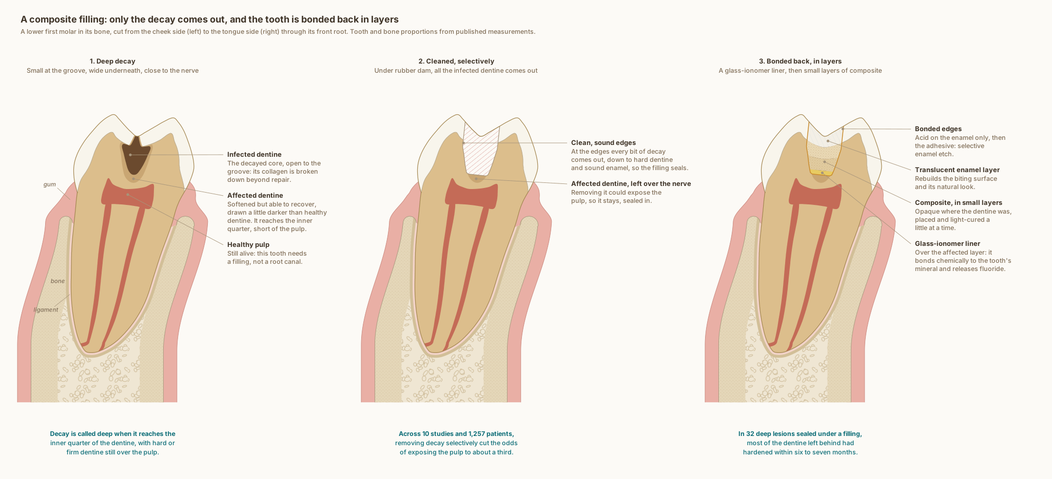
The same lower first molar in every panel, cut from the cheek side to the tongue side through its front root, with the gum, ligament and bone around it. The tooth and bone are drawn to published measurements; the decay and the filling show what typically happens, not one patient's tooth.
What the drawing is measured from
- Decay is called deep when it reaches the inner quarter of the dentine but still leaves a protective layer of hard or firm dentine over the pulp: the European Society of Endodontology's definition, restated in the 2026 S3-level guideline on deep caries from European dental societies. The guideline supports selective or stepwise removal over complete removal, to lower the risk of exposing the pulp, and with selective removal the dentine at the edges is still cleaned to hard, for a secure restoration. Schwendicke et al. 2026, Clinical Oral Investigations. DOI
- The decay is drawn as two zones, the two layers of carious dentine often called infected and affected dentine. In the outer layer the cross-links that hold the dentine's collagen together were irreversibly destroyed; in the inner layer the change was considered reversible. Kuboki, Ohgushi and Fusayama 1977, Journal of Dental Research, laboratory study. How far each zone reaches in the drawing is illustrative, not measured. DOI
- The decay's shape follows the textbook drawing of decay in a groove: small at the surface, widening toward the junction with the dentine, then wider in the dentine and on toward the pulp. Woelfel's Dental Anatomy, Figure 10-1.
- In deep lesions in teeth with healthy pulps, international consensus recommends selective removal to soft dentine: decayed dentine close to the pulp does not need to be removed, and the priorities are keeping healthy tooth tissue, achieving a seal and keeping the pulp healthy. Schwendicke et al. 2016, Advances in Dental Research, International Caries Consensus Collaboration. DOI
- A meta-analysis of 10 randomised trials with 1,257 patients found that leaving some decayed dentine over the pulp, in one visit or two, cut the odds of exposing the pulp to about a third of those with complete removal (odds ratio 0.31). Schwendicke, Dörfer and Paris 2013, Journal of Dental Research, systematic review and meta-analysis. DOI
- In 32 teeth with deep decay, the decay was only partly removed, then the teeth were lined with calcium hydroxide and sealed for six to seven months. When they were reopened the dentine was dry, it was hard in 80 per cent of the 30 teeth still in the study, and the bacteria in it had decreased significantly. Maltz et al. 2002, Quintessence International. PubMed
- The liner is glass ionomer, whose acid bonds chemically to the calcium of the tooth's mineral: in a laboratory study, about two-thirds of its acid groups were found bonded to hydroxyapatite. Yoshida et al. 2000, Journal of Dental Research. DOI
- Glass ionomers release fluoride and can take it up again, acting as a fluoride reservoir. Wiegand, Buchalla and Attin 2007, Dental Materials, review. DOI
- Acid on the enamel only, before a universal adhesive: in a randomised trial of 295 back-tooth fillings in 70 patients, fillings bonded this way showed significantly fewer defects at their edges after 24 months than those bonded with the adhesive alone. Barceleiro et al. 2023, Journal of Applied Oral Science. DOI
- The clinical steps are drawn as Dr. Khalid works: under rubber dam; all the infected dentine removed, with the dentine at the edges cleaned to hard and the enamel to sound so that the filling seals; a layer of affected dentine left over the pulp under a glass-ionomer liner; acid on the enamel only before the adhesive; and the composite built in small layers, opaque where the dentine was and translucent where the enamel was. The layer left over the pulp, about 0.7 mm at its deepest, and the 0.4 mm liner are representative thicknesses, not measurements.
- The enamel follows one measured dataset for this tooth, from cone-beam CT of 45 lower first molars: 1.58 mm at the front cheek-side cusp tip and 0.98 mm at the central fossa, its thinnest point. Zhang et al. 2026, BMC Oral Health. DOI
- Two canals in the front root, as drawn, are found in 94.4 per cent of lower first molars, and three in 2.3 per cent, from a systematic review of 41 studies covering 18,781 teeth. de Pablo et al. 2010, Journal of Endodontics. DOI
- Bone level: on cone-beam CT of 100 lower molar sites in 25 adults, the crest of the bone sat 2.03 to 2.11 mm below the enamel edge of first molars, with 1.14 to 1.19 mm of bone over the cheek side of the root near its neck. Othman 2026, The Saudi Dental Journal. DOI
- The cheek-side cortical plate near the crest is drawn from cone-beam CT of lower first and second molars, where it measured 1.1 to 1.4 mm. Sayed et al. 2021, The Saudi Dental Journal. DOI
- Further from the crest, on cone-beam CT of 100 people, between the lower second premolar and first molar: the cheek-side plate measured 1.93 mm at 4 mm below the enamel edge and 2.36 mm at 6 mm, and the tongue-side plate 2.18 and 2.09 mm. Gupta et al. 2025, BMC Oral Health. DOI
- The gum is drawn 1.2 mm thick, within the 0.8 to 1.5 mm measured by ultrasound across lower teeth in 200 periodontally healthy men. Eger, Müller and Heinecke 1996, Journal of Clinical Periodontology. DOI
- The tooth follows the textbook descriptions of a lower first molar rather than a symmetrical outline: the crown leans toward the tongue on its root, the front root's tip lies under the front cheek-side cusp, the roof of the pulp chamber sits at the level of the enamel edge with the floor in the root trunk, and the ligament around the root is drawn 0.2 mm wide, under the quarter of a millimetre the textbooks give, inside a thin lining of compact bone. Wheeler's Dental Anatomy, Physiology and Occlusion, 11th edition, and Woelfel's Dental Anatomy.
- The roof of the pulp chamber sits at the level of the enamel edge, where it lay in 97 per cent of 100 lower molars measured on radiographs, and the drawing's distance from the cheek-side cusp tip to that roof, 6.36 mm, is the study's mean. Deutsch and Musikant 2004, Journal of Endodontics. DOI
Use this diagram
Free to use, including commercially, with credit.
This diagram is published under CC BY 4.0. You may reproduce it in articles, teaching material, presentations and patient information, print or online, without asking. The one condition is that you credit Dr. Khalid Aletaibi and link back to this page.
It is published this way on purpose. The measurement in this drawing is not something the people selling the treatment tend to publish, so it is more useful spread than kept.
Credit line
A composite filling: only the decay comes out, and the tooth is bonded back in layers. Dr. Khalid Aletaibi, CC BY 4.0. https://khalidaletaibi.com/en/diagrams/composite-filling-steps
HTML embed
<figure>
  <img src="https://khalidaletaibi.com/figures/composite-filling-steps-en.svg" alt="A composite filling: only the decay comes out, and the tooth is bonded back in layers" width="100%">
  <figcaption>
    A composite filling: only the decay comes out, and the tooth is bonded back in layers. <a href="https://khalidaletaibi.com/en/diagrams/composite-filling-steps">Dr. Khalid Aletaibi</a>,
    <a href="https://creativecommons.org/licenses/by/4.0/">CC BY 4.0</a>
  </figcaption>
</figure>