Skip to content
Dr. Khalid AletaibiConservative Dentistry · Dubai

BIOMIMETIC RESTORATIONS

Bridges, only when nothing more conservative will work.

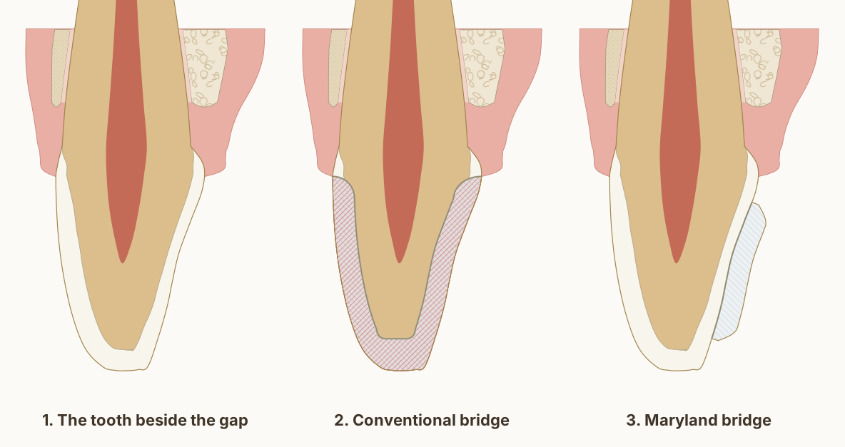
Three stages for the tooth beside a missing upper lateral incisor: the healthy central incisor, the same tooth cut down for a conventional bridge crown with the removed tooth hatched, and a Maryland bridge wing bonded to its palatal enamel with nothing cut.

A conventional bridge replaces one missing tooth by cutting down the two healthy teeth on either side. The technology works, well-made bridges can last fifteen years or more, but the trade is enormous, and it is no longer the only conservative answer to a single missing tooth.

Dr. Khalid AletaibiWritten by Dr. Khalid Aletaibi, DHA-licensed dentist

QUICK ANSWER

A conventional fixed bridge anchors an artificial replacement tooth (a pontic) to crowns cemented onto the teeth on either side of the gap. Modern systematic reviews report 5-year survival rates around 93%, dropping into the high 80s by ten years. The real question is rarely whether bridges work, they do. It is whether the two adjacent teeth genuinely need to be crowned at all, because once they are cut down, that decision cannot be reversed.

Visits

Usually 2

Anaesthetic

Local

Teeth prepared

Two neighbours

Reversibility

Permanent

Longevity

~93% at 5 yrs

What a bridge actually costs, two teeth, not one

A conventional bridge replaces a single missing tooth by anchoring a pontic to two abutment crowns on the teeth on either side of the gap. To make room for those crowns, the dentist removes the same circumferential band of enamel and dentine that a single crown requires, typically 0.5 to 2 mm of tissue all the way around, depending on the material (monolithic zirconia needs the least), but the cost is now paid by two teeth instead of one. Edelhoff and Sorensen quantified exactly how much sound tooth structure different preparation designs sacrifice [4], and a bridge preparation doubles every number in their tables.

The biological cost is doubled in the same way. Vital teeth that are crowned have a measurable risk of pulpal complications afterwards, and the Bergenholtz and Nyman study, now four decades old but still cited because the principle has not changed, documented the rate of pulp necrosis after prosthetic preparation in vital teeth [5]. When that risk is multiplied by two abutments, the cumulative probability of needing a root canal somewhere on the bridge over its lifetime is not negligible. None of this is an argument that bridges are wrong. It is an argument that the gap should be looked at very honestly before two healthy teeth are committed to filling it.

A lower second premolar and second molar beside a missing first molar, cut lengthways along the arch and seen from the cheek, with the gum and bone. The healed ridge is lower and narrower than before the extraction. Both neighbours are prepared with a 0.5 mm chamfer at the gum line, losing about 40 per cent of each crown in this section and keeping about 1.5 mm of dentine over the living pulp. The zirconia bridge joins two crowns to the false tooth through connectors about 3 mm tall, with rounded openings beneath them for cleaning.

Diagram page and reuse terms: A bridge for a missing back tooth: the two neighbours carry it

Sources and measurements
  • Drawn as Dr. Khalid works: a zirconia bridge for the back of the mouth, with a chamfer at the gum line on both neighbouring teeth, as on his crowns. The preparations are drawn with a 0.5 mm chamfer, 1.0 mm off the biting surface and walls converging 6 degrees each. The connectors are as tall as these crowns allow between the marginal ridges and the margins, 3.3 mm measured off the drawing. The false tooth's shape depends on the case; one rounded design is drawn. The second molar's biting surface, enamel and pulp chamber are the site's measured first-molar section, re-proportioned to the second molar.
  • In a laboratory comparison on model teeth, crown preparations made to each material's guidelines removed 21 per cent of a molar's crown for monolithic zirconia and 35 per cent for lithium disilicate, and 42 against 59 per cent of an incisor's. Schwindling et al. 2019, Clinical Oral Investigations. DOI 2, A bridge for a missing back tooth: the two neighbours carry it
  • A digital study of 720 crown preparation designs found that the margin's height and the chamfer's depth decide most of the tooth removed, and that materials which can be made thinner, such as monolithic zirconia, need less removed. Seidel et al. 2025, Clinical Oral Investigations. DOI 3, A bridge for a missing back tooth: the two neighbours carry it
  • In a clinical study of 20 three-unit zirconia bridges for back teeth, made with connectors of at least 9 mm² in cross-section, 15 were rated perfect in every respect after a mean of 31 months and five showed minor chipping of the veneering porcelain. Raigrodski et al. 2006, The Journal of Prosthetic Dentistry. DOI 4, A bridge for a missing back tooth: the two neighbours carry it
  • In laboratory tests of zirconia bridge frameworks, every framework broke at a connector, and connectors of at least 4 mm were recommended for long spans or where molars are replaced. Larsson et al. 2007, Journal of Oral Rehabilitation. DOI 5, A bridge for a missing back tooth: the two neighbours carry it
  • A systematic review of 29 laboratory and computer-model studies found that larger or taller connectors often made zirconia bridges more resistant to fracture, though connector area alone did not explain every result. Babiuc et al. 2026, Materials. DOI 6, A bridge for a missing back tooth: the two neighbours carry it
  • A systematic review of 20 studies found that six months after an extraction the ridge had lost 3.79 mm of width and 1.24 mm of height on the cheek side, and 0.84 and 0.80 mm beside the neighbouring teeth, most of it in the first three to six months. Tan et al. 2012, Clinical Oral Implants Research. DOI 7, A bridge for a missing back tooth: the two neighbours carry it
  • Cone-beam CT of 219 sites under the false teeth of implant bridges, three years after they were fitted, measured the gum between the false tooth and the bone at 2.32 mm in the upper jaw and 2.20 mm in the lower. Pozzi et al. 2015, Clinical Implant Dentistry and Related Research. DOI 8, A bridge for a missing back tooth: the two neighbours carry it
  • The teeth follow Wheeler's measurements and drawings: the lower second premolar's crown 8.0 mm, root 14.5 mm, 7.0 mm wide and 5.0 mm at the cervix; the lower second molar's crown 7.0 mm, root 13.0 mm, 10.5 mm wide and 8.0 mm at the cervix; the missing first molar 11.0 mm wide. Their outlines are traced from Wheeler's buccal drawings. Nelson, Wheeler's Dental Anatomy, Physiology and Occlusion, 11th edition, Tables 10.2, 12.1 and 12.2, Figures 10.13 and 12.18.
  • Central sections of 40 lower second premolars measured the enamel at the contacts at 1.35 to 1.40 mm on the mesial side and 1.44 to 1.46 mm on the distal. Fernandes et al. 2011, Brazilian Oral Research. DOI 10, A bridge for a missing back tooth: the two neighbours carry it
  • Bone level: on cone-beam CT of 100 lower molar sites in 25 adults, the crest of the bone sat 2.03 to 2.11 mm below the enamel edge of first molars, with 1.14 to 1.19 mm of bone over the cheek side of the root near its neck. Othman 2026, The Saudi Dental Journal. DOI 11, A bridge for a missing back tooth: the two neighbours carry it
  • The roof of the pulp chamber sits at the level of the enamel edge, where it lay in 97 per cent of 100 lower molars measured on radiographs, and the drawing's distance from the cheek-side cusp tip to that roof, 6.36 mm, is the study's mean. Deutsch and Musikant 2004, Journal of Endodontics. DOI 12, A bridge for a missing back tooth: the two neighbours carry it

A close-up of the joint between the premolar's zirconia crown and the false tooth in the step-by-step figure, with a 1 mm scale. The connector measures 3.3 mm tall here, as much as these crowns allow above the margins; beneath it a rounded opening leaves room for floss and small brushes, and the crown's margin sits at the gum.

Diagram page and reuse terms: The connector

Sources and measurements
  • Drawn from the bridge in the step-by-step figure: the connector's height is measured off the drawing, 3.3 mm at its thinnest, and the opening beneath it is drawn round. The cement is drawn 0.1 mm thick.
  • In a clinical study of 20 three-unit zirconia bridges for back teeth, made with connectors of at least 9 mm² in cross-section, 15 were rated perfect in every respect after a mean of 31 months and five showed minor chipping of the veneering porcelain. Raigrodski et al. 2006, The Journal of Prosthetic Dentistry. DOI 2, The connector
  • In laboratory tests of zirconia bridge frameworks, every framework broke at a connector, and connectors of at least 4 mm were recommended for long spans or where molars are replaced. Larsson et al. 2007, Journal of Oral Rehabilitation. DOI 3, The connector
  • A systematic review of 29 laboratory and computer-model studies found that larger or taller connectors often made zirconia bridges more resistant to fracture, though connector area alone did not explain every result. Babiuc et al. 2026, Materials. DOI 4, The connector

A close-up under the false tooth in the step-by-step figure, with a 1 mm scale. Its rounded underside rests lightly on the gum without pressing into it; the gum over the healed ridge is drawn 2.2 mm thick, and the underside curves up at each side, leaving room for floss and small brushes.

Diagram page and reuse terms: Under the false tooth

Sources and measurements
  • Drawn from the bridge in the step-by-step figure. The false tooth's shape depends on the case; one rounded design is drawn, touching the gum over about 3 mm and rising away from it at each side.
  • Cone-beam CT of 219 sites under the false teeth of implant bridges, three years after they were fitted, measured the gum between the false tooth and the bone at 2.32 mm in the upper jaw and 2.20 mm in the lower. Pozzi et al. 2015, Clinical Implant Dentistry and Related Research. DOI 2, Under the false tooth
  • A systematic review of 20 studies found that six months after an extraction the ridge had lost 3.79 mm of width and 1.24 mm of height on the cheek side, and 0.84 and 0.80 mm beside the neighbouring teeth, most of it in the first three to six months. Tan et al. 2012, Clinical Oral Implants Research. DOI 3, Under the false tooth

What the longevity data shows

When the indication for a conventional bridge is correct and the construction is precise, the survival data is reasonably good. The Pjetursson 2007 systematic review compared tooth-supported fixed dental prostheses to implant-supported alternatives and reported 5-year survival rates around 93% for conventional FDPs [1]. The Pjetursson 2004 systematic review of fixed partial dentures with at least 5 years of follow-up is the foundational dataset for that finding and remains the most cited body of evidence on the subject [2]. A more recent meta-analysis specifically on zirconia-based tooth-supported FDPs reported broadly similar short-term survival figures, with the contemporary all-ceramic era now reaching the maturity required for confident recommendations [3].

Two findings from the same body of literature deserve to be read alongside the survival numbers, because they tell you what actually fails. The first is that the bridge framework itself is rarely the failure point. The dominant failure modes in the long-term studies are recurrent decay at the abutment margins, loss of pulp vitality in the abutment teeth, and, eventually, loss of one of the abutment teeth itself, which usually means the entire bridge has to be replaced or converted to something larger. A bridge does not just put two teeth at risk for the cost of one: it links their fates, so that the failure of either one ends the restoration.

The upper central incisor next to the gap, cut from the lip side to the palate side, with its gum and bone. For a conventional bridge it is cut down for a lithium disilicate crown with a chamfer at the gum line, losing over 40 per cent of this section, and the canine on the other side of the gap is crowned too. For a Maryland bridge one ceramic wing is bonded to its palatal enamel and nothing is cut.

Diagram page and reuse terms: A bridge for a missing front tooth: what each design takes from the tooth beside the gap

Sources and measurements
  • Drawn as Dr. Khalid works: for a conventional bridge, a lithium disilicate crown with a chamfer at the gum line, drawn with 1.0 mm of reduction at the margin and on the walls and 1.5 mm at the edge; for a Maryland bridge, a single lithium disilicate wing on the central incisor with no preparation, drawn 1.0 mm thick and kept short of the gum and of the edge. Over 40 per cent is the share of this section the crown takes, measured off the drawing.
  • In a laboratory comparison on model teeth, crown preparations made to each material's guidelines removed 21 per cent of a molar's crown for monolithic zirconia and 35 per cent for lithium disilicate, and 42 against 59 per cent of an incisor's. Schwindling et al. 2019, Clinical Oral Investigations. DOI 2, A bridge for a missing front tooth: what each design takes from the tooth beside the gap
  • A systematic review of 2,300 resin-bonded bridges found survival of 91.4 per cent at five years and 82.9 per cent at ten; bridges held by one wing survived longer and came loose less often than those held by two. Thoma et al. 2017, Clinical Oral Implants Research. DOI 3, A bridge for a missing front tooth: what each design takes from the tooth beside the gap
  • Of 310 single-wing zirconia bridges replacing incisors, followed for a mean of about seven years, 97.3 per cent were estimated to survive 15 years; 17 came loose, and 16 of them were bonded back. Kern et al. 2025, The Journal of Prosthetic Dentistry. DOI 4, A bridge for a missing front tooth: what each design takes from the tooth beside the gap
  • Labial enamel thickness at 1, 3 and 5 mm above the cementoenamel junction: 0.48, 0.81 and 0.95 mm. Cone-beam CT of 1,600 maxillary central incisors in patients aged 18 to 40, Diagnostics 2024, PMC11592583. In agreement with micro-CT of 20 central incisors, which found a range of 0.461 to 0.966 mm. Enamel also thins significantly with age, so these are not lifetime values. DOI 5, A bridge for a missing front tooth: what each design takes from the tooth beside the gap
  • Pulp distances measured on cone-beam CT across 200 teeth: palatal fossa to the pulp chamber 2.21 mm, incisal edge to the pulp 5.02 mm. Kohok et al. 2025, PMID 41193532. PubMed 6, A bridge for a missing front tooth: what each design takes from the tooth beside the gap
  • The tooth follows Wheeler's measurements for the upper central incisor: crown 10.5 mm, root 13.0 mm, 7.0 mm labiolingually at the crown and 6.0 mm at the cervix, with the crests of contour in the cervical third. Nelson, Wheeler's Dental Anatomy, Physiology and Occlusion, 11th edition, Table 6.1.
  • At the upper central incisors of 88 periodontally healthy adults, the gum overlapped the enamel by 1.41 mm, the bone crest lay 2.05 mm from the cementoenamel junction, the free gingiva was 1.12 mm thick at its base and the bone 0.45 mm thick at the crest. Alpiste-Illueca 2004, The International Journal of Periodontics and Restorative Dentistry. PubMed 8, A bridge for a missing front tooth: what each design takes from the tooth beside the gap
  • Cone-beam CT of 120 people measured the labial bone over upper central incisors at 0.73, 0.69 and 0.60 mm in the coronal, middle and apical thirds, with the palatal bone significantly thicker. AlTarawneh et al. 2018, Journal of Prosthodontics. DOI 9, A bridge for a missing front tooth: what each design takes from the tooth beside the gap

How I decide whether a conventional bridge is the right answer

I work down a conservative ladder before I consider cutting two healthy teeth for a bridge. If the gap is in a position where it does not affect chewing, speech, aesthetics, or the stability of the neighbouring teeth, the most conservative answer is sometimes to leave it alone and monitor. If the gap matters, an implant is almost always the more biologically conservative option, it replaces one tooth with one root, without touching anything else. And if the adjacent teeth are completely intact and the gap is in the anterior region, a resin-bonded cantilever bridge is often the right answer: Kern's long-term data shows fifteen-year survival of anterior all-ceramic cantilever resin-bonded FDPs in the very high range, with almost no preparation on the supporting tooth [6].

A conventional crown-retained bridge is the right answer in a narrow set of situations: when an implant is genuinely contraindicated, insufficient bone, medical contraindications, or the patient's informed preference after a full conversation about the alternatives. AND the adjacent teeth already need crowns for independent reasons. When both conditions are true, replacing the missing tooth with a pontic between those crowns is efficient and conservative, because the preparation needed to happen anyway. When only one condition is true, the conservative answer is almost always something else.

Three upper front teeth drawn to Wheeler's measurements, crowns down as in the mouth. From the front only the false lateral incisor shows; the wing is hidden behind the central incisor, drawn dashed. From the palate the wing, the connector and the false tooth are one piece: the wing covers most of the central's palatal enamel, clear of the gum, the edge and the other contact, and the canine is not touched.

Diagram page and reuse terms: A Maryland bridge from the front and from the palate

Sources and measurements
  • Drawn as Dr. Khalid makes a Maryland bridge: one wing, on the central incisor, with no preparation, in lithium disilicate. The wing is drawn from 2.6 mm below the cervical line to 1.5 mm short of the edge, at least 1 mm from the gum and 0.8 mm from the other contact; the connector sits at the contact, clear of the false tooth's edge. The false tooth's shape depends on the case. The gum is drawn 1.0 mm past each cervical line, as in the site's other figures.
  • A systematic review of 2,300 resin-bonded bridges found survival of 91.4 per cent at five years and 82.9 per cent at ten; bridges held by one wing survived longer and came loose less often than those held by two. Thoma et al. 2017, Clinical Oral Implants Research. DOI 2, A Maryland bridge from the front and from the palate
  • Of 310 single-wing zirconia bridges replacing incisors, followed for a mean of about seven years, 97.3 per cent were estimated to survive 15 years; 17 came loose, and 16 of them were bonded back. Kern et al. 2025, The Journal of Prosthetic Dentistry. DOI 3, A Maryland bridge from the front and from the palate
  • The upper central incisor, lateral incisor and canine follow Wheeler's measurements, crowns 10.5, 9.0 and 10.0 mm long and 8.5, 6.5 and 7.5 mm wide, with their outlines traced from Wheeler's labial and lingual drawings. Nelson, Wheeler's Dental Anatomy, Physiology and Occlusion, 11th edition, Tables 6.1, 6.2 and 8.1, Figures 6.2, 6.3, 6.13, 6.14, 8.2 and 8.3.

A close-up of the wing in the step-by-step figure, with a 0.5 mm scale. Nothing is cut: the tooth's own palatal enamel is etched, and the ceramic wing, its inner surface sandblasted, etched, cleaned and primed with silane, is bonded to it with resin cement.

Diagram page and reuse terms: The wing on untouched enamel

Sources and measurements
  • Drawn as Dr. Khalid bonds a Maryland bridge: no preparation; the enamel etched; the ceramic sandblasted, etched with hydrofluoric acid, cleaned with phosphoric acid and primed with silane, as for all his ceramics. The cement is drawn 0.1 mm thick, and the etched layers wider than life so they can be seen; their widths are not measurements.
  • In a laboratory study on the ground enamel of 45 upper central incisors, phosphoric acid dissolved from about 3.5 micrometres of enamel (5 per cent acid for 15 seconds) to 27 micrometres (37 per cent acid for 60 seconds), so in life the etched band is far thinner than a drawing can show. Legler, Retief and Bradley 1990, American Journal of Orthodontics and Dentofacial Orthopedics. DOI 2, The wing on untouched enamel
  • In a laboratory study on 70 upper central incisors, ceramic veneers bonded to an all-enamel surface held about twice the shear bond strength of those bonded to dentine alone, about 20 against 10 MPa, and keeping at least 40 per cent of the bonding surface in enamel preserved the bond. Zhu et al. 2022, BMC Oral Health. DOI 3, The wing on untouched enamel
  • In 62 lithium disilicate veneers in 12 patients, measured on silicone replicas by micro-CT, the median gap at the margin was 100 micrometres without preparation and 140 micrometres with minimal preparation, and the internal gap averaged about 0.17 to 0.22 mm. Karagözoğlu, Toksavul and Toman 2016, Quintessence International. DOI 4, The wing on untouched enamel
  • Of 310 single-wing zirconia bridges replacing incisors, followed for a mean of about seven years, 97.3 per cent were estimated to survive 15 years; 17 came loose, and 16 of them were bonded back. Kern et al. 2025, The Journal of Prosthetic Dentistry. DOI 5, The wing on untouched enamel
  • Labial enamel thickness at 1, 3 and 5 mm above the cementoenamel junction: 0.48, 0.81 and 0.95 mm. Cone-beam CT of 1,600 maxillary central incisors in patients aged 18 to 40, Diagnostics 2024, PMC11592583. In agreement with micro-CT of 20 central incisors, which found a range of 0.461 to 0.966 mm. Enamel also thins significantly with age, so these are not lifetime values. DOI 6, The wing on untouched enamel

A close-up through the false lateral incisor, with a 1 mm scale. Its rounded base rests 1 mm into a shallow hollow of the gum, so it seems to come out of the gum like a natural tooth, and the gum keeps about 2 mm over the bone. Its exact shape depends on the case.

Diagram page and reuse terms: The false tooth on the gum

Sources and measurements
  • The false tooth is the site's measured incisor crown scaled to Wheeler's lateral incisor, 9.0 mm long and 6.0 mm from lip to palate; its base is drawn rounded, 1 mm into the gum, with 2 mm of gum over the bone. The false tooth's shape depends on the case; one rounded design is drawn.
  • Cone-beam CT of 219 sites under the false teeth of implant bridges, three years after they were fitted, measured the gum between the false tooth and the bone at 2.32 mm in the upper jaw and 2.20 mm in the lower. Pozzi et al. 2015, Clinical Implant Dentistry and Related Research. DOI 2, The false tooth on the gum
  • At 36 false-tooth sites of single-wing zirconia bridges replacing incisors, followed for 4 to more than 10 years, the gum stayed stable and even grew a little at its margin, giving the false tooth an emergence like a natural tooth's. Türp et al. 2025, Journal of Esthetic and Restorative Dentistry. DOI 3, The false tooth on the gum
  • A systematic review of 20 studies found that six months after an extraction the ridge had lost 3.79 mm of width and 1.24 mm of height on the cheek side, and 0.84 and 0.80 mm beside the neighbouring teeth, most of it in the first three to six months. Tan et al. 2012, Clinical Oral Implants Research. DOI 4, The false tooth on the gum
  • The upper central incisor, lateral incisor and canine follow Wheeler's measurements, crowns 10.5, 9.0 and 10.0 mm long and 8.5, 6.5 and 7.5 mm wide, with their outlines traced from Wheeler's labial and lingual drawings. Nelson, Wheeler's Dental Anatomy, Physiology and Occlusion, 11th edition, Tables 6.1, 6.2 and 8.1, Figures 6.2, 6.3, 6.13, 6.14, 8.2 and 8.3.

What happens, step by step

  1. 1

    We weigh the alternatives first

    Before any tooth is touched we check whether an implant, a resin-bonded bridge, or simply monitoring the gap would protect more of your healthy teeth.

  2. 2

    Prepare the two abutment teeth

    If a conventional bridge is the right answer, we reduce the two teeth either side of the gap by about 0.5 to 2 mm all round, depending on the material, to make room for the supporting crowns.

  3. 3

    Impression and temporary

    We take a digital or physical impression and fit a temporary bridge to protect the prepared teeth while the laboratory builds the final one.

  4. 4

    Fit and bond the bridge

    We check the fit, bite, and shade, then seal the bridge in place with an adhesive or conventional cement chosen to guard the margins against leakage.

  5. 5

    Review and maintenance

    We review the abutment margins and gum health over time, since the long-term outcome depends on keeping those two teeth decay-free.

Survival when a conventional bridge is the right choice
Conventional bridge survival at 5 years[1]93%

Pooled 5-year survival from systematic reviews of tooth-supported fixed bridges. Survival does not answer whether the two neighbouring teeth needed to be prepared in the first place.

Conservative profile

  • How invasiveHigh
  • Healthy teeth preservedLow
  • Replaces the tooth without surgeryHigh
  • ReversibilityLow

COMMON QUESTIONS

What patients ask most.

What's the difference between a bridge and an implant?
An implant replaces one root with one root, a titanium fixture is placed into the bone of the missing tooth, and a crown is placed on top of it. The neighbouring teeth are not touched. A conventional bridge does the opposite: it leaves the gap in the bone empty and cuts down the two adjacent teeth so that crowns can be placed on them and a pontic suspended between them. Whenever bone allows it, the implant is the more biologically conservative option because it spares the adjacent teeth entirely.
How long does a bridge last?
In current systematic reviews, well-made conventional bridges show 5-year survival rates around 93%, dropping into the high 80s by ten years and into the high 70s by fifteen years. The bridge framework itself is rarely the failure point, the dominant failure modes are recurrent decay at one of the abutment margins, loss of vitality in an abutment tooth, or eventual loss of an abutment tooth that ends the entire restoration. Those supporting teeth are what your routine visits look after: at your check-up and cleaning, set at 4, 6 or 12 months for you, the margins and the space under the bridge are checked and cleaned, and you are shown how to clean beneath it at home. What happens at a cleaning is set out in your cleaning, step by step.
What is a Maryland or resin-bonded bridge?
A resin-bonded bridge replaces a missing tooth by bonding a thin extension (a 'wing') onto the back of an adjacent tooth, instead of cementing a full crown over it. Almost no preparation is needed on the supporting tooth. The long-term data for the modern single-retainer cantilever design, particularly in the anterior region, is excellent, and it is often the most conservative option for replacing an anterior tooth when the supporting tooth is intact and an implant is not yet possible.
Can I just leave the gap?
Sometimes that is the right answer. Not every missing tooth has to be replaced, particularly second molars at the very back, where the absence often does not affect chewing efficiency or the stability of the rest of the mouth. The risks of leaving a gap are tooth drift, opposing tooth super-eruption, food impaction, and altered chewing patterns. Whether those risks are clinically meaningful depends on the specific gap. We assess and recommend honestly rather than reflexively filling every space.
Can a bridge be replaced with an implant later?
Sometimes, but not always. If the abutment teeth are still healthy when the bridge eventually fails, the bridge can be removed and the missing-tooth site can be replaced with an implant, leaving the previously prepared abutments to receive single crowns. But if the bridge has been on the teeth for ten or fifteen years, one or both abutments have often developed decay or pulp problems by that point, and the situation becomes considerably more complex. This is one of the reasons to choose carefully at the start.
How is a bridge cemented?
Adhesive bonding under proper isolation when the materials and remaining tooth structure allow it, conventional cementation when they do not. The cementation step is not a passive 'gluing in': it determines how well the abutment teeth are sealed against bacterial leakage at the margins, which is the most common pathway to long-term failure. For lithium disilicate and full-ceramic bridges in particular, an adhesive bonding protocol is part of why the modern survival numbers are as good as they are.

Speak to us

Would you prefer we call you?

Leave your name and number and Dr. Khalid's team will call you back to answer your questions and find a time that suits you.

Bridge, implant, or Maryland, the answer depends on your specific gap.

We will assess the missing-tooth site, the adjacent teeth, the bone, and your own preferences, then explain which of the three options the evidence supports for your specific situation, and why.

Book a Consultation

← Biomimetic Restorations & Endodontics

Call Dr. Khalid directlyDiscuss your concerns and questions