Skip to content
Dr. Khalid AletaibiConservative Dentistry · Dubai

BIOMIMETIC RESTORATIONS

Root canal treatment, only when it's truly needed.

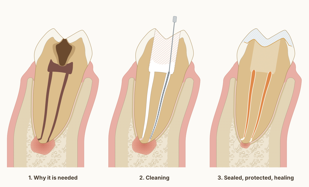
Three stages of root canal treatment in a lower molar: an infected pulp with inflamed bone at the root tip, then the canals cleaned and shaped, then the canals filled and the tooth sealed and covered while the bone heals.

A root canal is a tooth-saving procedure, not a tooth-replacing one. That makes it the conservative option by definition: keeping your own root is almost always better than removing it and replacing it with something manufactured. The most important question I ask isn't how to do it well, it's whether the tooth needs one at all. When the answer is yes, the second question is how to do it while preserving the maximum amount of healthy dentine.

Dr. Khalid AletaibiWritten by Dr. Khalid Aletaibi, DHA-licensed dentist

QUICK ANSWER

Root canal treatment removes inflamed or infected pulp tissue from inside a tooth, disinfects the canal system, and seals it. Done minimally invasively, the access cavity is kept as small as the anatomy allows, because every millimetre of dentine left around the canals is what stops the tooth fracturing later. In current systematic reviews, modern non-surgical root canal treatment is associated with tooth survival rates above 85% at 8-10 years when followed by an adequate coronal restoration. The single most important predictor of long-term success is not the obturation technique, it is the quality of the final restoration that seals the access.

Visits

1 to 2

Anaesthetic

Local

Goal

Saves the tooth

Discomfort

Minimal

Longevity

>85% at 8-10 yrs

What a root canal actually is, and what it isn't

When the pulp inside a tooth becomes irreversibly inflamed or infected, most often from deep caries that has reached the nerve, sometimes from trauma or a deep crack, the body cannot heal it on its own. The two options are removing the entire tooth or removing only the pulp tissue and saving the tooth. Modern endodontic treatment, what many people call having the nerve taken out, is the second option: a careful, microscope-assisted cleaning and disinfection of the internal canal system, followed by a sealed obturation and a definitive bonded restoration.

The outcome data on properly performed non-surgical root canal treatment is genuinely good. A systematic review of tooth survival after root canal treatment reported pooled survival rates above 85% at 8-10 years across many studies [1]. Earlier systematic reviews of treatment outcome, looking specifically at periapical healing rather than tooth retention, show success rates in the same range when modern technique is used [2]. These are not numbers from idealised laboratory conditions. They are real-world outcomes from clinical practice.

The same lower first molar in every panel, cut from the cheek side to the tongue side through its front root, with the gum, ligament and bone around it. The tooth and bone are drawn to published measurements; the three stages show what typically happens, not one patient's tooth.

Diagram page and reuse terms: Root canal treatment: cleaned, sealed, protected, and the bone can heal

Sources and measurements
  • Two canals in the front root, as drawn, are found in 94.4 per cent of lower first molars, and three in 2.3 per cent, from a systematic review of 41 studies covering 18,781 teeth. de Pablo et al. 2010, Journal of Endodontics. DOI 1, Root canal treatment: cleaned, sealed, protected, and the bone can heal
  • The inflammation at the root tip comes from inside the tooth: apical periodontitis is a chronic inflammation of the tissues around the root tip caused by agents of endodontic origin, above all infection inside the root canal. Nair 2006, International Endodontic Journal, review. DOI 2, Root canal treatment: cleaned, sealed, protected, and the bone can heal
  • The note under the second panel comes from a laboratory study of 42 extracted upper second premolars, loaded without breaking them: the root canal procedures themselves reduced the tooth's relative stiffness by 5 per cent, an occlusal cavity by 20 per cent, and a cavity through both walls facing the neighbouring teeth by 63 per cent. Reeh, Messer and Douglas 1989, Journal of Endodontics. DOI 3, Root canal treatment: cleaned, sealed, protected, and the bone can heal
  • The note under the third panel: of 126 roots treated with an inflamed tip and followed yearly for up to four years, 111 (88 per cent) showed signs of healing. Healing had begun within the first year in at least 89 per cent of those, and complete healing sometimes took four years. Ørstavik 1996, International Endodontic Journal. DOI 4, Root canal treatment: cleaned, sealed, protected, and the bone can heal
  • Where the canal exits: across 1,331 roots, the main exit of the canal lay a mean 0.80 mm from the root tip in lower molars, always within 1 mm, and sat exactly at the tip in only 40 per cent of teeth; when it lay to one side, the cheek side was the commonest. Martos et al. 2009, International Endodontic Journal. DOI 5, Root canal treatment: cleaned, sealed, protected, and the bone can heal
  • The narrowest point of the canal: in micro-CT scans of 56 extracted molars, in canals with a single narrowing, it lay a mean 0.76 mm inside the canal's exit. Hu et al. 2026, BMC Oral Health. DOI 6, Root canal treatment: cleaned, sealed, protected, and the bone can heal
  • The root filling is drawn as Dr. Khalid places it: a single gutta-percha cone that ends at the canal's narrowest point, in a bioceramic sealer that fills the canal beyond it to the exit. In 307 teeth filled this way in a private practice and followed for an average of 30 months, overall success was 90.9 per cent; sealer passed beyond the root tip in 47.4 per cent of them, with no significant effect on the outcome. Chybowski et al. 2018, Journal of Endodontics, retrospective study. DOI 7, Root canal treatment: cleaned, sealed, protected, and the bone can heal
  • Bioceramic (hydraulic calcium silicate) sealers were compared with other sealer types across 55 clinical reports covering 5,913 teeth: success rates were comparable, with no increase in pain after treatment. Donnermeyer et al. 2026, Journal of Dentistry, systematic review and meta-analysis. DOI 8, Root canal treatment: cleaned, sealed, protected, and the bone can heal
  • In dogs' teeth with induced lesions at the root tip, a bioceramic sealer pushed beyond the tip on purpose produced less inflammation and more new bone at four months than a resin-based calcium silicate sealer. Elsayed et al. 2025, Clinical Oral Investigations, animal study. DOI 9, Root canal treatment: cleaned, sealed, protected, and the bone can heal
  • Bone level: on cone-beam CT of 100 lower molar sites in 25 adults, the crest of the bone sat 2.03 to 2.11 mm below the enamel edge of first molars, with 1.14 to 1.19 mm of bone over the cheek side of the root near its neck. Othman 2026, The Saudi Dental Journal. DOI 10, Root canal treatment: cleaned, sealed, protected, and the bone can heal
  • Bone beyond the root tip: on high-resolution cone-beam CT of 1,400 teeth, the lower first molar had 4.45 mm of bone from the exit of the cheek-side front canal to the cheek-side cortical plate, and 5.63 mm from the exit of the tongue-side front canal to the tongue-side plate. Porto et al. 2020, Journal of Applied Oral Science. DOI 11, Root canal treatment: cleaned, sealed, protected, and the bone can heal
  • The cheek-side cortical plate near the crest is drawn from cone-beam CT of lower first and second molars, where it measured 1.1 to 1.4 mm. Sayed et al. 2021, The Saudi Dental Journal. DOI 12, Root canal treatment: cleaned, sealed, protected, and the bone can heal
  • Further from the crest, on cone-beam CT of 100 people, between the lower second premolar and first molar: the cheek-side plate measured 1.93 mm at 4 mm below the enamel edge and 2.36 mm at 6 mm, and the tongue-side plate 2.18 and 2.09 mm. Gupta et al. 2025, BMC Oral Health. DOI 13, Root canal treatment: cleaned, sealed, protected, and the bone can heal
  • The gum is drawn 1.2 mm thick, within the 0.8 to 1.5 mm measured by ultrasound across lower teeth in 200 periodontally healthy men. Eger, Müller and Heinecke 1996, Journal of Clinical Periodontology. DOI 14, Root canal treatment: cleaned, sealed, protected, and the bone can heal
  • The tooth follows the textbook descriptions of a lower first molar rather than a symmetrical outline: the crown leans toward the tongue on its root, the front root's tip lies under the front cheek-side cusp, the roof of the pulp chamber sits at the level of the enamel edge with the floor in the root trunk, and the ligament around the root is drawn 0.2 mm wide, under the quarter of a millimetre the textbooks give, inside a thin lining of compact bone. Wheeler's Dental Anatomy, Physiology and Occlusion, 11th edition, and Woelfel's Dental Anatomy.
  • Two choices in the drawing are clinical rather than measured, and both are drawn as Dr. Khalid works. The opening removes the whole roof of the pulp chamber, leaving no ledge over the pulp horns, so every canal entrance lies in view; its walls flare slightly and stop short of the cusp tips. The tooth is then rebuilt with bonded composite placed directly over the canal entrances, filling the chamber and the opening, and a bonded overlay covering the cusps.
  • The roof of the pulp chamber sits at the level of the enamel edge, where it lay in 97 per cent of 100 lower molars measured on radiographs, and the drawing's distance from the cheek-side cusp tip to that roof, 6.36 mm, is the study's mean. Deutsch and Musikant 2004, Journal of Endodontics. DOI 17, Root canal treatment: cleaned, sealed, protected, and the bone can heal

What I do to avoid a root canal in the first place

For decades, deep caries close to the pulp meant a root canal was on the table. The evidence has moved on. The 2016 international consensus on managing carious lesions recommends leaving a thin layer of softer affected dentine over the pulp in deep cavities and sealing it under a well-bonded restoration, rather than excavating until the pulp is exposed [6]. In practice this single change has prevented an enormous number of root canals worldwide, simply by stopping clinicians from creating the very pulp exposure they were trying to avoid.

When the pulp is already symptomatic, vital pulp therapy is the next level of conservative management. A 2019 systematic review of full pulpotomy in mature carious teeth with symptoms of irreversible pulpitis reported clinical success rates well above 80% in selected cases [5]. That is a substantial body of evidence supporting an option that, twenty years ago, would have been dismissed outright. It does not replace root canal treatment for every case, but it expands the conservative menu meaningfully, and for the right tooth in the right patient it preserves a vital pulp that would otherwise have been removed.

When a root canal is the right answer: how I do it differently

There are situations where a root canal is unambiguously the right call: a frankly necrotic pulp, a periapical lesion on radiograph, irreversible symptoms unresponsive to conservative care. When that is the case, modern endodontics emphasises preserving as much pericervical dentine as possible. The Clark and Khademi paper on modern molar access advanced the principle that the access cavity should be as small as possible while still allowing the canals to be located and instrumented [4]. Larger straight-line access used to be standard teaching. The trade-off, convenience for the operator at the cost of long-term tooth strength, is no longer one I am willing to make.

The other principle that has shaped modern endodontics is older than I am: the long-term outcome of a root-treated tooth depends on the quality of the final restoration at least as much as it depends on the obturation. Ray and Trope's classic 1995 study showed exactly this, teeth with both a good root filling and a good coronal restoration fared dramatically better than teeth with one or the other alone [3]. The implication is unambiguous: the day the canal is sealed, the access cavity must be sealed permanently with a properly bonded restoration. A temporary filling left in place for weeks is the most common cause of preventable endodontic failure I see in second-opinion cases.

A close-up of the front root's tip from the same measured molar as the step-by-step figure, with a 1 mm scale. Each canal narrows just before it exits, usually a little to one side of the tip, and that exit is where bacteria and their toxins reach the bone.

Diagram page and reuse terms: Where the canal opens into the bone

Sources and measurements
  • Where the canal exits: across 1,331 roots, the main exit of the canal lay a mean 0.80 mm from the root tip in lower molars, always within 1 mm, and sat exactly at the tip in only 40 per cent of teeth; when it lay to one side, the cheek side was the commonest. Martos et al. 2009, International Endodontic Journal. DOI 1, Where the canal opens into the bone
  • The narrowest point of the canal: in micro-CT scans of 56 extracted molars, in canals with a single narrowing, it lay a mean 0.76 mm inside the canal's exit. Hu et al. 2026, BMC Oral Health. DOI 2, Where the canal opens into the bone
  • The inflammation at the root tip comes from inside the tooth: apical periodontitis is a chronic inflammation of the tissues around the root tip caused by agents of endodontic origin, above all infection inside the root canal. Nair 2006, International Endodontic Journal, review. DOI 3, Where the canal opens into the bone
  • The ligament that holds the root in its socket is drawn 0.2 mm wide, under the quarter of a millimetre the textbook gives, and the socket is lined by a thin layer of compact bone, the lamina dura, which shows on an X-ray as a white line around the root. Woelfel's Dental Anatomy.
  • The size and outline of the inflamed area are representative rather than measured from a study, so they carry no number. On an X-ray it shows as a dark area around the root tip, because the inflamed tissue has replaced bone.

Looking down through the opening of the same measured molar, with the whole roof of the chamber removed so that no ledge hides an entrance. Three canals is the commonest arrangement, two in the front root and one behind; about a third of these teeth have a fourth.

Diagram page and reuse terms: The floor of the pulp chamber

Sources and measurements
  • The canal count comes from a systematic review of 41 studies covering 18,781 lower first molars: three canals in 61.3 per cent and four in 35.7 per cent; the front root held two canals in 94.4 per cent, and the back root a single canal in 62.7 per cent. de Pablo et al. 2010, Journal of Endodontics. DOI 1, The floor of the pulp chamber
  • The entrances sit where the textbook places them in this tooth: the cheek-side front entrance close to the front cheek-side cusp tip, the tongue-side front entrance in a more central position rather than under its cusp, and a single back canal's large entrance just behind the centre of the crown. Woelfel's Dental Anatomy.
  • Finding them follows the laws for locating pulp chambers and canal entrances that Krasner and Rankow proposed after examining 500 pulp chambers of extracted teeth. Krasner and Rankow 2004, Journal of Endodontics. DOI 3, The floor of the pulp chamber
  • The opening removes the whole roof of the chamber, leaving no ledge over the pulp horns, so that every entrance lies in view. Poor access design and missed canals are among the recognised causes of inflammation at the root tip that persists after treatment. Nair 2006, International Endodontic Journal, review. DOI 4, The floor of the pulp chamber

What happens, step by step

  1. 1

    Diagnosis and numbing

    We confirm the pulp is genuinely involved and fully numb the tooth, so the procedure is comfortable throughout.

  2. 2

    Cleaning and disinfection

    Under magnification, the inflamed or infected pulp is removed and the canal system is carefully disinfected.

  3. 3

    Sealing the canals

    Each cleaned canal is filled with a single gutta-percha cone in a bioceramic sealer, which fills the canal to its very end and sets with the canal's own moisture, sealing it against re-infection.

  4. 4

    A definitive bonded restoration

    A well-bonded restoration seals the access, the single biggest predictor of long-term success.

Outcomes with modern technique
Tooth survival at 8-10 years[1]85%
Full pulpotomy success, selected cases[5]80%

Pooled rates from current systematic reviews; both figures are reported as "above" these values with a well-sealed final restoration.

Conservative profile

  • Saves the natural toothHigh
  • Discomfort (modern technique)Low
  • Long-term survivalHigh

COMMON QUESTIONS

What patients ask most.

Is root canal treatment painful?
With modern anaesthesia and microscope-assisted technique, the procedure itself is essentially painless. Most discomfort patients expect to feel comes from the inflamed tooth before the appointment, not from the treatment itself. A systematic review of 72 studies found pain in about 81% of patients before treatment, 40% a day after it and 11% a week after, with its severity falling to minimal levels within seven days.[7] Some mild tenderness in the days after is normal as the tissues around the root settle. If a patient has significant pain after a root canal, that is a clinical signal worth investigating, not a normal expectation.
Is a root canal risky?
It is a routine, well-studied treatment, and its risks are mostly small and temporary. Tenderness for a few days afterwards is common. Less often, the area flares up with swelling, a canal turns out to be hidden or calcified, or a crack that was not visible before becomes the limiting factor; rarely, a fine instrument separates inside a canal. Over the long term, pooled data show 86% to 93% of root-treated teeth still in place 2 to 10 years later, and protecting the tooth afterwards, with a crown in that review, was the factor most strongly linked to keeping it.[1] The larger risk is usually the alternative: an infected tooth left untreated can spread infection into the surrounding bone and is eventually lost. Each of these complications, how often it happens and what lowers the risk are set out in the real risks of a root canal.
How long does a root canal take?
Usually one or two appointments. Most of the time goes on finding, cleaning and sealing every canal, so a front tooth with a single canal is quicker than a molar with three or four, and for a molar a single appointment of up to about two hours is not unusual. When there is active infection or swelling, a medicated dressing between two visits can be the better plan. A Cochrane review of 47 studies found no difference in healing between finishing in one visit and in several, although more people reported pain during the first week after single-visit treatment.[8]
Is it better to extract the tooth and replace it with an implant?
Almost never, when the tooth is restorable. A natural tooth, even one that has been root-treated, has a periodontal ligament, proprioception, and a biological connection to the bone that no implant can replicate. Modern endodontic outcomes are excellent, and saving the natural tooth is the conservative answer in nearly every case where it is technically feasible. Extraction is reserved for teeth that cannot be reasonably restored, not for teeth where root canal treatment is the right call. The comparison itself is set out in full on can this tooth be saved.
How long does a root-treated tooth last?
In current systematic reviews, tooth survival after non-surgical root canal treatment is well above 85% at 8-10 years when followed by an adequate coronal restoration. Many root-treated teeth function for the rest of the patient's life. The dominant predictor of long-term survival is the quality of the final restoration that seals the access cavity and protects the remaining tooth structure, not the obturation technique alone. So that seal is worth looking after: at your check-up and cleaning, set at 4, 6 or 12 months for you, the edges of the final restoration are checked and cleaned, so new decay or a gap at the edge is found early, while it is still small. What happens at a cleaning is set out in your cleaning, step by step.
Do I always need a crown after a root canal?
Not always. The reflexive 'root canal equals crown' rule is being re-examined by current evidence. For a tooth with a small access cavity and intact walls, a well-bonded direct or indirect restoration can be the right answer; for a tooth with significant tissue loss and undermined cusps, a crown or onlay is genuinely needed. The decision is made tooth by tooth based on how much structure is left, not by reflex. Where a molar does need covering, an endocrown takes its retention from the pulp chamber instead of a post drilled into the root.
What if my tooth is sensitive but I'm not sure I need a root canal?
Sensitivity is not the same as irreversible pulpitis. A careful examination and a properly performed pulp test are what separate a tooth that needs a root canal from one that needs a small filling, a desensitising treatment, or simply observation. A second opinion before agreeing to a root canal, particularly when the tooth is currently functional and only mildly symptomatic, is one of the most conservative things a patient can do.
Can a root canal fail? What happens then?
Yes, a small percentage do, most often because of a missed canal, persistent infection, or a coronal seal that broke down. The first response is not extraction. Re-treatment (cleaning and re-sealing the canal system) or, less often, a microsurgical apicoectomy can save a tooth that initially failed. Each option has a real evidence base behind it, and an honest conversation about which one is appropriate is part of the work.

Speak to us

Would you prefer we call you?

Leave your name and number and Dr. Khalid's team will call you back to answer your questions and find a time that suits you.

Told you need a root canal? Get a second opinion first.

Sometimes the answer is yes, and the procedure is the right call. Sometimes a more conservative option is available and was missed. We'll examine the tooth carefully and tell you exactly what the evidence supports for your specific situation.

Book a Consultation

← Biomimetic Restorations & Endodontics

Call Dr. Khalid directlyDiscuss your concerns and questions