Measured diagram
What is inside a front tooth rebuilt with bonding?
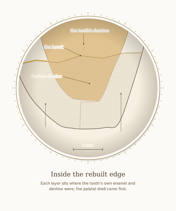
A close-up of the rebuilt edge from the step-by-step figure, with a 1 mm scale. The palatal shell came first, formed against the silicone key where the tooth's palatal enamel was; the dentine shades follow the tooth's own dentine; and the front enamel layer completes the front and the edge. The dashed line is the break they replace.
What the drawing is measured from
- The rebuild follows two principles from one textbook. The additive principle: in a direct restoration the etching and bonding are extended beyond the defect onto intact enamel, to blend and reinforce the restoration; its example is a small chipped edge restored without preparation: the tooth was etched, bonded and restored well beyond the fracture line under rubber dam. And natural guided stratification: a silicone index made from a wax-up or mock-up holds a palatal shell of enamel-like material, dentine-like increments are placed where the natural dentine was, and the last increment is the labial enamel. Magne and Belser 2022, Biomimetic Restorative Dentistry, volume 1, pages 319, 330 and 331.
- Drawn as Dr. Khalid layers a chipped edge: a palatal shell against a silicone key made from a wax-up or mock-up of the agreed shape, then dentine shades where the tooth's dentine was, then a front enamel layer. The shell follows the drawing's own palatal enamel, as no published thickness exists for it, and the level and line of the break are illustrative.
- Labial enamel thickness at 1, 3 and 5 mm above the cementoenamel junction: 0.48, 0.81 and 0.95 mm. Cone-beam CT of 1,600 maxillary central incisors in patients aged 18 to 40, Diagnostics 2024, PMC11592583. In agreement with micro-CT of 20 central incisors, which found a range of 0.461 to 0.966 mm. Enamel also thins significantly with age, so these are not lifetime values. DOI
- Two things on this drawing are anatomically representative but are not taken from published measurements, and are therefore not labelled with a number: the palatal enamel profile, and the width of the pulp space. Only the labelled values are sourced.
Use this diagram
Free to use, including commercially, with credit.
This diagram is published under CC BY 4.0. You may reproduce it in articles, teaching material, presentations and patient information, print or online, without asking. The one condition is that you credit Dr. Khalid Aletaibi and link back to this page.
It is published this way on purpose. The measurement in this drawing is not something the people selling the treatment tend to publish, so it is more useful spread than kept.
Credit line
Inside the rebuilt edge. Dr. Khalid Aletaibi, CC BY 4.0. https://khalidaletaibi.com/en/diagrams/bond-inside
HTML embed
<figure>
  <img src="https://khalidaletaibi.com/figures/bond-inside-en.svg" alt="Inside the rebuilt edge" width="100%">
  <figcaption>
    Inside the rebuilt edge. <a href="https://khalidaletaibi.com/en/diagrams/bond-inside">Dr. Khalid Aletaibi</a>,
    <a href="https://creativecommons.org/licenses/by/4.0/">CC BY 4.0</a>
  </figcaption>
</figure>