Diagram library
Measured dental diagrams, free to reuse.
Four clinical drawings, each built to measured anatomy rather than sketched, with the studies every dimension comes from printed underneath. Published under CC BY 4.0, so you may use them anywhere with credit.
CC BY 4.0, credit Dr. Khalid Aletaibi. Reuse terms
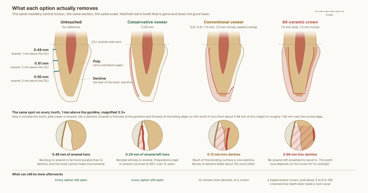
How much tooth structure does a veneer remove compared with a crown?
The same upper central incisor in every panel, one midline section, drawn to scale from measured anatomy. Hatched red is tissue that does not grow back.
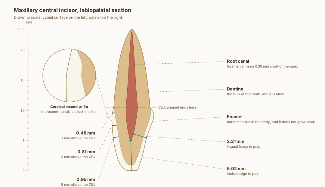
How thick is the enamel on an upper front tooth?
Drawn to scale from published measurements, with every labelled dimension measured back off the drawn geometry rather than asserted beside it. The enamel really is that thin at the neck.
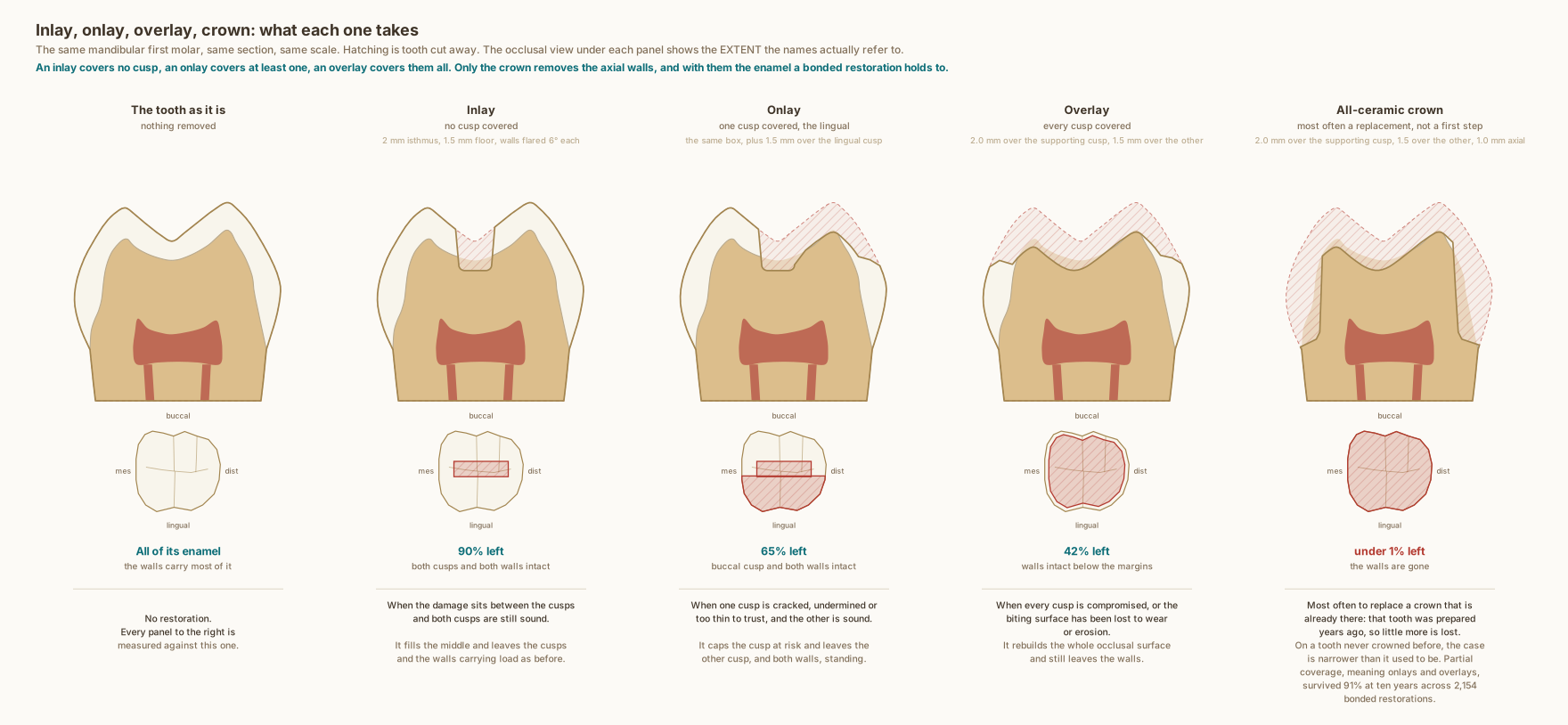
What is the difference between an inlay, an onlay, an overlay and a crown?
The same mandibular first molar in every panel, drawn to scale from measured anatomy. The percentages are areas measured off these drawings by scanline every 0.005 millimetres, within this one section, and are not whole-tooth volumes.
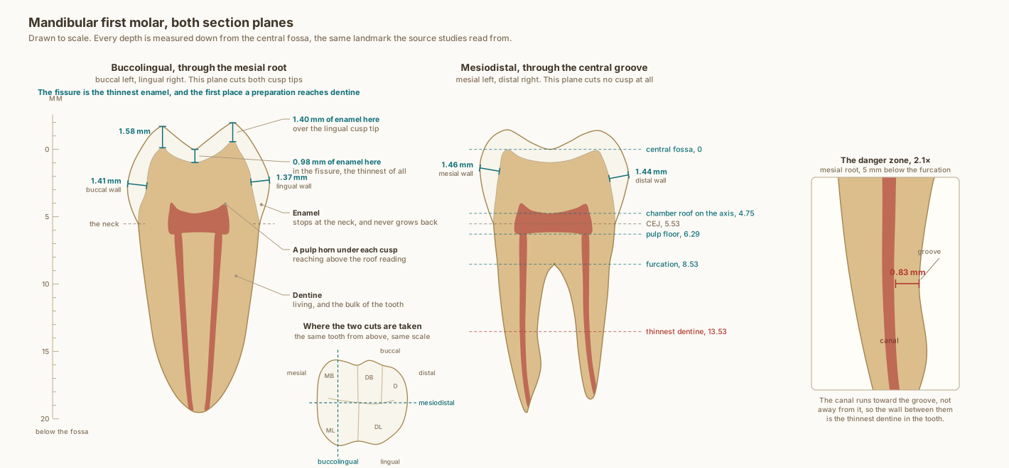
What does a lower first molar look like in cross section?
Drawn to scale, with every depth measured down from the central fossa, which is the landmark the source studies read from. Each stated dimension is measured back off the drawn geometry rather than asserted beside it.