Measured diagram
How is a chipped front tooth rebuilt with bonding, and does any of the tooth have to be cut?
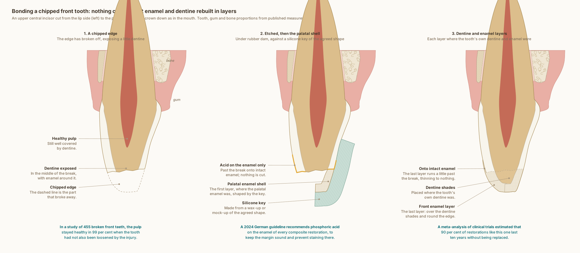
The same upper central incisor in every panel, cut from the lip side to the palate side, with the gum, ligament and bone around it. The tooth and the tissues at its neck are drawn to published measurements; the break, the etched enamel, the key and the layers show what typically happens, not one patient's tooth.
What the drawing is measured from
- The rebuild follows two principles from one textbook. The additive principle: in a direct restoration the etching and bonding are extended beyond the defect onto intact enamel, to blend and reinforce the restoration; its example is a small chipped edge restored without preparation: the tooth was etched, bonded and restored well beyond the fracture line under rubber dam. And natural guided stratification: a silicone index made from a wax-up or mock-up holds a palatal shell of enamel-like material, dentine-like increments are placed where the natural dentine was, and the last increment is the labial enamel. Magne and Belser 2022, Biomimetic Restorative Dentistry, volume 1, pages 319, 330 and 331.
- In 455 crown-fractured permanent incisors followed for a mean of 2.3 years and up to 17, the pulp survived in 99 per cent of teeth the injury had not also loosened or displaced, and in none of them did it die; when the tooth had also been loosened, pulp death in fractures through enamel and dentine, like this one, rose from 0 to 28 per cent. Robertson et al. 2000, International Journal of Paediatric Dentistry. DOI
- A 2024 German clinical guideline on direct composite restorations, agreed by strong consensus, recommends etching the enamel of every direct composite restoration with phosphoric acid, to improve the long-term quality of the enamel margin and prevent discoloration there. It finds both rubber dam and other isolation workable, with rubber dam possibly helping restorations last longer. These process recommendations are consensus-based. Sekundo et al. 2024, The Journal of Adhesive Dentistry, part 2. DOI
- Acid on the enamel only, before a universal adhesive: in a randomised trial of 295 back-tooth fillings in 70 patients, fillings bonded this way showed significantly fewer defects at their edges after 24 months than those bonded with the adhesive alone. Barceleiro et al. 2023, Journal of Applied Oral Science. DOI
- In fillings at the necks of teeth, a meta-analysis of 10 randomised trials found less staining and fewer adaptation defects at the edges when the enamel was etched selectively before a self-etch adhesive, and fewer fillings lost at three years. Szesz et al. 2016, Journal of Dentistry, systematic review and meta-analysis. DOI
- A Cochrane review of six randomised trials with 1,342 participants found low-certainty evidence that rubber dam may lower the failure rate of fillings compared with cotton rolls at six months, and very uncertain evidence at other times; all the trials were at high risk of bias, and the results came from fillings at the necks of teeth and in children's back teeth. Miao et al. 2021, Cochrane Database of Systematic Reviews. DOI
- A meta-analysis of 21 prospective clinical trials of front-tooth composites estimated that 90 per cent of Class IV restorations, which rebuild a broken corner or edge of a front tooth, last 10 years without being replaced; bulk fracture was the main reason for replacement, more often with microfilled composites. Twelve of the trials started before 1991. Heintze, Rousson and Hickel 2015, Dental Materials. DOI
- A critical appraisal of clinical and laboratory publications: a bevel on the enamel margin was traditionally placed for retention and a gradual colour transition, but modern adhesives, composites and technique make adequate front-tooth composites possible without one, with a more conservative preparation, a smaller restored surface and more tooth kept when the restoration is redone. Baratieri and Ritter 2005, Journal of Esthetic and Restorative Dentistry. DOI
- The steps are drawn as Dr. Khalid works: nothing is cut, not even a bevel; the work is done under rubber dam; phosphoric acid goes on the enamel only, past the break, before the adhesive; a silicone key made from a wax-up or mock-up of the agreed shape guides the palatal enamel shell; then dentine shades go where the tooth's own dentine was, and a last enamel layer rebuilds the front and runs a little onto intact enamel. The level and line of the break are illustrative, the shell follows the drawing's palatal enamel, and the layers are drawn to the tooth's own enamel and dentine.
- Labial enamel thickness at 1, 3 and 5 mm above the cementoenamel junction: 0.48, 0.81 and 0.95 mm. Cone-beam CT of 1,600 maxillary central incisors in patients aged 18 to 40, Diagnostics 2024, PMC11592583. In agreement with micro-CT of 20 central incisors, which found a range of 0.461 to 0.966 mm. Enamel also thins significantly with age, so these are not lifetime values. DOI
- Pulp distances measured on cone-beam CT across 200 teeth: palatal fossa to the pulp chamber 2.21 mm, incisal edge to the pulp 5.02 mm. Kohok et al. 2025, PMID 41193532. PubMed
- Two things on this drawing are anatomically representative but are not taken from published measurements, and are therefore not labelled with a number: the palatal enamel profile, and the width of the pulp space. Only the labelled values are sourced.
- The tooth follows Wheeler's measurements for the upper central incisor: crown 10.5 mm, root 13.0 mm, 7.0 mm labiolingually at the crown and 6.0 mm at the cervix, with the crests of contour in the cervical third. Nelson, Wheeler's Dental Anatomy, Physiology and Occlusion, 11th edition, Table 6.1.
- At the upper central incisors of 88 periodontally healthy adults, the gum overlapped the enamel by 1.41 mm, the bone crest lay 2.05 mm from the cementoenamel junction, the free gingiva was 1.12 mm thick at its base and the bone 0.45 mm thick at the crest. Alpiste-Illueca 2004, The International Journal of Periodontics and Restorative Dentistry. PubMed
- Cone-beam CT of 120 people measured the labial bone over upper central incisors at 0.73, 0.69 and 0.60 mm in the coronal, middle and apical thirds, with the palatal bone significantly thicker. AlTarawneh et al. 2018, Journal of Prosthodontics. DOI
Use this diagram
Free to use, including commercially, with credit.
This diagram is published under CC BY 4.0. You may reproduce it in articles, teaching material, presentations and patient information, print or online, without asking. The one condition is that you credit Dr. Khalid Aletaibi and link back to this page.
It is published this way on purpose. The measurement in this drawing is not something the people selling the treatment tend to publish, so it is more useful spread than kept.
Credit line
Bonding a chipped front tooth: nothing cut, the lost enamel and dentine rebuilt in layers. Dr. Khalid Aletaibi, CC BY 4.0. https://khalidaletaibi.com/en/diagrams/bond-steps
HTML embed
<figure>
  <img src="https://khalidaletaibi.com/figures/bond-steps-en.svg" alt="Bonding a chipped front tooth: nothing cut, the lost enamel and dentine rebuilt in layers" width="100%">
  <figcaption>
    Bonding a chipped front tooth: nothing cut, the lost enamel and dentine rebuilt in layers. <a href="https://khalidaletaibi.com/en/diagrams/bond-steps">Dr. Khalid Aletaibi</a>,
    <a href="https://creativecommons.org/licenses/by/4.0/">CC BY 4.0</a>
  </figcaption>
</figure>