Measured diagram
What does a bridge for a missing back tooth involve, and how much of the neighbouring teeth is removed?
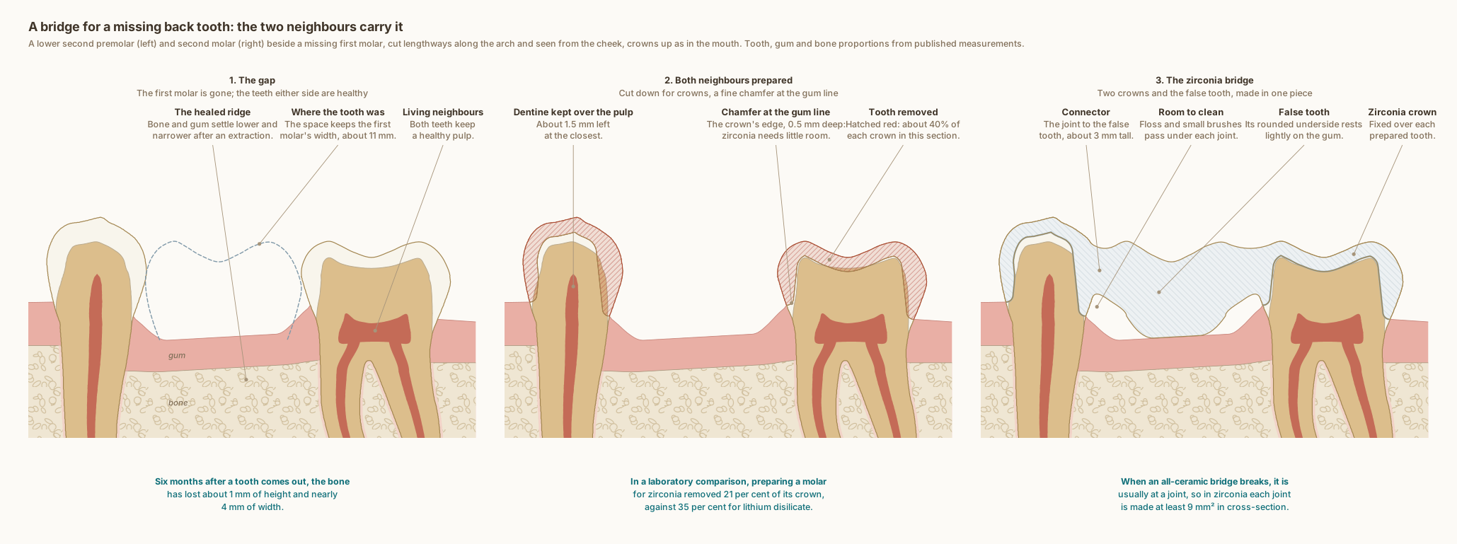
A lower second premolar and second molar beside a missing first molar, cut lengthways along the arch and seen from the cheek, with the gum and bone. The healed ridge is lower and narrower than before the extraction. Both neighbours are prepared with a 0.5 mm chamfer at the gum line, losing about 40 per cent of each crown in this section and keeping about 1.5 mm of dentine over the living pulp. The zirconia bridge joins two crowns to the false tooth through connectors about 3 mm tall, with rounded openings beneath them for cleaning.
What the drawing is measured from
- Drawn as Dr. Khalid works: a zirconia bridge for the back of the mouth, with a chamfer at the gum line on both neighbouring teeth, as on his crowns. The preparations are drawn with a 0.5 mm chamfer, 1.0 mm off the biting surface and walls converging 6 degrees each. The connectors are as tall as these crowns allow between the marginal ridges and the margins, 3.3 mm measured off the drawing. The false tooth's shape depends on the case; one rounded design is drawn. The second molar's biting surface, enamel and pulp chamber are the site's measured first-molar section, re-proportioned to the second molar.
- In a laboratory comparison on model teeth, crown preparations made to each material's guidelines removed 21 per cent of a molar's crown for monolithic zirconia and 35 per cent for lithium disilicate, and 42 against 59 per cent of an incisor's. Schwindling et al. 2019, Clinical Oral Investigations. DOI
- A digital study of 720 crown preparation designs found that the margin's height and the chamfer's depth decide most of the tooth removed, and that materials which can be made thinner, such as monolithic zirconia, need less removed. Seidel et al. 2025, Clinical Oral Investigations. DOI
- In a clinical study of 20 three-unit zirconia bridges for back teeth, made with connectors of at least 9 mm² in cross-section, 15 were rated perfect in every respect after a mean of 31 months and five showed minor chipping of the veneering porcelain. Raigrodski et al. 2006, The Journal of Prosthetic Dentistry. DOI
- In laboratory tests of zirconia bridge frameworks, every framework broke at a connector, and connectors of at least 4 mm were recommended for long spans or where molars are replaced. Larsson et al. 2007, Journal of Oral Rehabilitation. DOI
- A systematic review of 29 laboratory and computer-model studies found that larger or taller connectors often made zirconia bridges more resistant to fracture, though connector area alone did not explain every result. Babiuc et al. 2026, Materials. DOI
- A systematic review of 20 studies found that six months after an extraction the ridge had lost 3.79 mm of width and 1.24 mm of height on the cheek side, and 0.84 and 0.80 mm beside the neighbouring teeth, most of it in the first three to six months. Tan et al. 2012, Clinical Oral Implants Research. DOI
- Cone-beam CT of 219 sites under the false teeth of implant bridges, three years after they were fitted, measured the gum between the false tooth and the bone at 2.32 mm in the upper jaw and 2.20 mm in the lower. Pozzi et al. 2015, Clinical Implant Dentistry and Related Research. DOI
- The teeth follow Wheeler's measurements and drawings: the lower second premolar's crown 8.0 mm, root 14.5 mm, 7.0 mm wide and 5.0 mm at the cervix; the lower second molar's crown 7.0 mm, root 13.0 mm, 10.5 mm wide and 8.0 mm at the cervix; the missing first molar 11.0 mm wide. Their outlines are traced from Wheeler's buccal drawings. Nelson, Wheeler's Dental Anatomy, Physiology and Occlusion, 11th edition, Tables 10.2, 12.1 and 12.2, Figures 10.13 and 12.18.
- Central sections of 40 lower second premolars measured the enamel at the contacts at 1.35 to 1.40 mm on the mesial side and 1.44 to 1.46 mm on the distal. Fernandes et al. 2011, Brazilian Oral Research. DOI
- Bone level: on cone-beam CT of 100 lower molar sites in 25 adults, the crest of the bone sat 2.03 to 2.11 mm below the enamel edge of first molars, with 1.14 to 1.19 mm of bone over the cheek side of the root near its neck. Othman 2026, The Saudi Dental Journal. DOI
- The roof of the pulp chamber sits at the level of the enamel edge, where it lay in 97 per cent of 100 lower molars measured on radiographs, and the drawing's distance from the cheek-side cusp tip to that roof, 6.36 mm, is the study's mean. Deutsch and Musikant 2004, Journal of Endodontics. DOI
Use this diagram
Free to use, including commercially, with credit.
This diagram is published under CC BY 4.0. You may reproduce it in articles, teaching material, presentations and patient information, print or online, without asking. The one condition is that you credit Dr. Khalid Aletaibi and link back to this page.
It is published this way on purpose. The measurement in this drawing is not something the people selling the treatment tend to publish, so it is more useful spread than kept.
Credit line
A bridge for a missing back tooth: the two neighbours carry it. Dr. Khalid Aletaibi, CC BY 4.0. https://khalidaletaibi.com/en/diagrams/bridge-back-steps
HTML embed
<figure>
  <img src="https://khalidaletaibi.com/figures/bridge-back-steps-en.svg" alt="A bridge for a missing back tooth: the two neighbours carry it" width="100%">
  <figcaption>
    A bridge for a missing back tooth: the two neighbours carry it. <a href="https://khalidaletaibi.com/en/diagrams/bridge-back-steps">Dr. Khalid Aletaibi</a>,
    <a href="https://creativecommons.org/licenses/by/4.0/">CC BY 4.0</a>
  </figcaption>
</figure>