Measured diagram
How much of the neighbouring tooth does a bridge for a missing front tooth take?
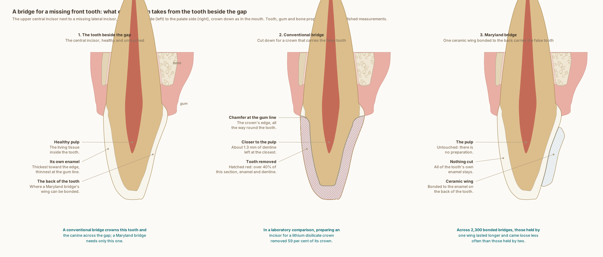
The upper central incisor next to the gap, cut from the lip side to the palate side, with its gum and bone. For a conventional bridge it is cut down for a lithium disilicate crown with a chamfer at the gum line, losing over 40 per cent of this section, and the canine on the other side of the gap is crowned too. For a Maryland bridge one ceramic wing is bonded to its palatal enamel and nothing is cut.
What the drawing is measured from
- Drawn as Dr. Khalid works: for a conventional bridge, a lithium disilicate crown with a chamfer at the gum line, drawn with 1.0 mm of reduction at the margin and on the walls and 1.5 mm at the edge; for a Maryland bridge, a single lithium disilicate wing on the central incisor with no preparation, drawn 1.0 mm thick and kept short of the gum and of the edge. Over 40 per cent is the share of this section the crown takes, measured off the drawing.
- In a laboratory comparison on model teeth, crown preparations made to each material's guidelines removed 21 per cent of a molar's crown for monolithic zirconia and 35 per cent for lithium disilicate, and 42 against 59 per cent of an incisor's. Schwindling et al. 2019, Clinical Oral Investigations. DOI
- A systematic review of 2,300 resin-bonded bridges found survival of 91.4 per cent at five years and 82.9 per cent at ten; bridges held by one wing survived longer and came loose less often than those held by two. Thoma et al. 2017, Clinical Oral Implants Research. DOI
- Of 310 single-wing zirconia bridges replacing incisors, followed for a mean of about seven years, 97.3 per cent were estimated to survive 15 years; 17 came loose, and 16 of them were bonded back. Kern et al. 2025, The Journal of Prosthetic Dentistry. DOI
- Labial enamel thickness at 1, 3 and 5 mm above the cementoenamel junction: 0.48, 0.81 and 0.95 mm. Cone-beam CT of 1,600 maxillary central incisors in patients aged 18 to 40, Diagnostics 2024, PMC11592583. In agreement with micro-CT of 20 central incisors, which found a range of 0.461 to 0.966 mm. Enamel also thins significantly with age, so these are not lifetime values. DOI
- Pulp distances measured on cone-beam CT across 200 teeth: palatal fossa to the pulp chamber 2.21 mm, incisal edge to the pulp 5.02 mm. Kohok et al. 2025, PMID 41193532. PubMed
- The tooth follows Wheeler's measurements for the upper central incisor: crown 10.5 mm, root 13.0 mm, 7.0 mm labiolingually at the crown and 6.0 mm at the cervix, with the crests of contour in the cervical third. Nelson, Wheeler's Dental Anatomy, Physiology and Occlusion, 11th edition, Table 6.1.
- At the upper central incisors of 88 periodontally healthy adults, the gum overlapped the enamel by 1.41 mm, the bone crest lay 2.05 mm from the cementoenamel junction, the free gingiva was 1.12 mm thick at its base and the bone 0.45 mm thick at the crest. Alpiste-Illueca 2004, The International Journal of Periodontics and Restorative Dentistry. PubMed
- Cone-beam CT of 120 people measured the labial bone over upper central incisors at 0.73, 0.69 and 0.60 mm in the coronal, middle and apical thirds, with the palatal bone significantly thicker. AlTarawneh et al. 2018, Journal of Prosthodontics. DOI
Use this diagram
Free to use, including commercially, with credit.
This diagram is published under CC BY 4.0. You may reproduce it in articles, teaching material, presentations and patient information, print or online, without asking. The one condition is that you credit Dr. Khalid Aletaibi and link back to this page.
It is published this way on purpose. The measurement in this drawing is not something the people selling the treatment tend to publish, so it is more useful spread than kept.
Credit line
A bridge for a missing front tooth: what each design takes from the tooth beside the gap. Dr. Khalid Aletaibi, CC BY 4.0. https://khalidaletaibi.com/en/diagrams/bridge-front-steps
HTML embed
<figure>
  <img src="https://khalidaletaibi.com/figures/bridge-front-steps-en.svg" alt="A bridge for a missing front tooth: what each design takes from the tooth beside the gap" width="100%">
  <figcaption>
    A bridge for a missing front tooth: what each design takes from the tooth beside the gap. <a href="https://khalidaletaibi.com/en/diagrams/bridge-front-steps">Dr. Khalid Aletaibi</a>,
    <a href="https://creativecommons.org/licenses/by/4.0/">CC BY 4.0</a>
  </figcaption>
</figure>