Measured diagram
How is a Maryland bridge wing attached without cutting the tooth?
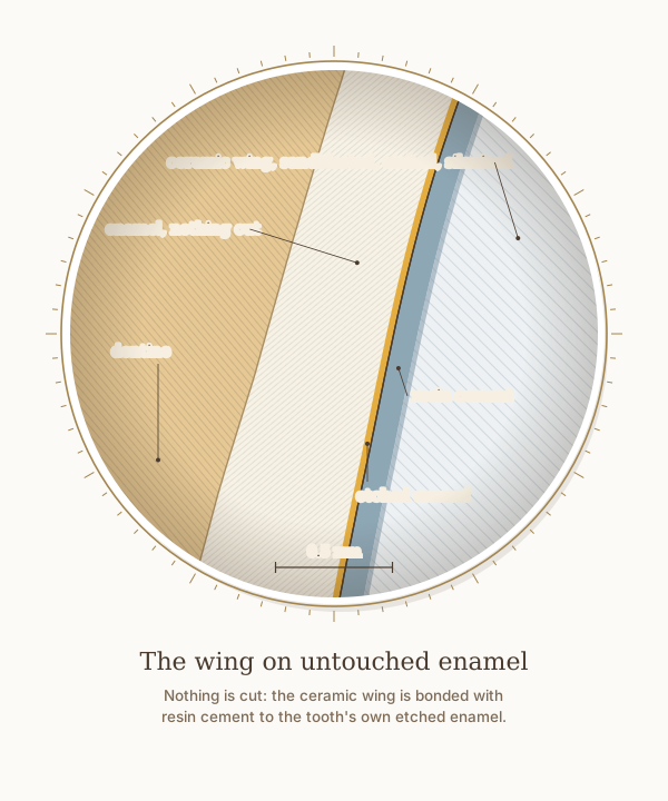
A close-up of the wing in the step-by-step figure, with a 0.5 mm scale. Nothing is cut: the tooth's own palatal enamel is etched, and the ceramic wing, its inner surface sandblasted, etched, cleaned and primed with silane, is bonded to it with resin cement.
What the drawing is measured from
- Drawn as Dr. Khalid bonds a Maryland bridge: no preparation; the enamel etched; the ceramic sandblasted, etched with hydrofluoric acid, cleaned with phosphoric acid and primed with silane, as for all his ceramics. The cement is drawn 0.1 mm thick, and the etched layers wider than life so they can be seen; their widths are not measurements.
- In a laboratory study on the ground enamel of 45 upper central incisors, phosphoric acid dissolved from about 3.5 micrometres of enamel (5 per cent acid for 15 seconds) to 27 micrometres (37 per cent acid for 60 seconds), so in life the etched band is far thinner than a drawing can show. Legler, Retief and Bradley 1990, American Journal of Orthodontics and Dentofacial Orthopedics. DOI
- In a laboratory study on 70 upper central incisors, ceramic veneers bonded to an all-enamel surface held about twice the shear bond strength of those bonded to dentine alone, about 20 against 10 MPa, and keeping at least 40 per cent of the bonding surface in enamel preserved the bond. Zhu et al. 2022, BMC Oral Health. DOI
- In 62 lithium disilicate veneers in 12 patients, measured on silicone replicas by micro-CT, the median gap at the margin was 100 micrometres without preparation and 140 micrometres with minimal preparation, and the internal gap averaged about 0.17 to 0.22 mm. Karagözoğlu, Toksavul and Toman 2016, Quintessence International. DOI
- Of 310 single-wing zirconia bridges replacing incisors, followed for a mean of about seven years, 97.3 per cent were estimated to survive 15 years; 17 came loose, and 16 of them were bonded back. Kern et al. 2025, The Journal of Prosthetic Dentistry. DOI
- Labial enamel thickness at 1, 3 and 5 mm above the cementoenamel junction: 0.48, 0.81 and 0.95 mm. Cone-beam CT of 1,600 maxillary central incisors in patients aged 18 to 40, Diagnostics 2024, PMC11592583. In agreement with micro-CT of 20 central incisors, which found a range of 0.461 to 0.966 mm. Enamel also thins significantly with age, so these are not lifetime values. DOI
Use this diagram
Free to use, including commercially, with credit.
This diagram is published under CC BY 4.0. You may reproduce it in articles, teaching material, presentations and patient information, print or online, without asking. The one condition is that you credit Dr. Khalid Aletaibi and link back to this page.
It is published this way on purpose. The measurement in this drawing is not something the people selling the treatment tend to publish, so it is more useful spread than kept.
Credit line
The wing on untouched enamel. Dr. Khalid Aletaibi, CC BY 4.0. https://khalidaletaibi.com/en/diagrams/bridge-wing
HTML embed
<figure>
  <img src="https://khalidaletaibi.com/figures/bridge-wing-en.svg" alt="The wing on untouched enamel" width="100%">
  <figcaption>
    The wing on untouched enamel. <a href="https://khalidaletaibi.com/en/diagrams/bridge-wing">Dr. Khalid Aletaibi</a>,
    <a href="https://creativecommons.org/licenses/by/4.0/">CC BY 4.0</a>
  </figcaption>
</figure>