Measured diagram
Can a tooth be kept alive when the decay has reached the nerve?
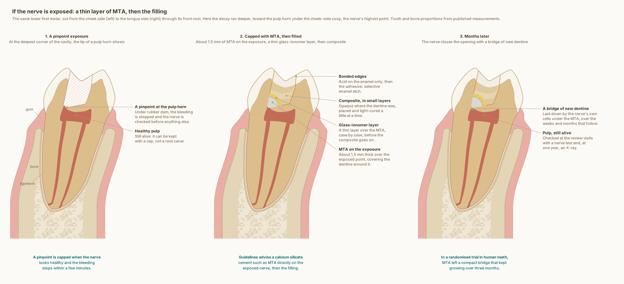
Three stages in the same measured lower molar. A pinpoint of the nerve shows at the deepest corner of the cavity, at the tip of a pulp horn. Under rubber dam, MTA goes directly on it, about 1.5 mm thick over the exposure and spread over the dentine around it, then a thin glass-ionomer layer and composite in small layers. Over the following weeks and months the nerve's own cells lay down a bridge of new dentine under the MTA, and the tooth is checked at review visits.
What the drawing is measured from
- Decay is called deep when it reaches the inner quarter of the dentine but still leaves a protective layer of hard or firm dentine over the pulp: the European Society of Endodontology's definition, restated in the 2026 S3-level guideline on deep caries from European dental societies. The guideline supports selective or stepwise removal over complete removal, to lower the risk of exposing the pulp, and with selective removal the dentine at the edges is still cleaned to hard, for a secure restoration. Schwendicke et al. 2026, Clinical Oral Investigations. DOI 1
- When the decay has reached the pulp, the European Society of Endodontology advises working under rubber dam, stopping the bleeding with sodium hypochlorite or chlorhexidine, placing a calcium silicate cement such as MTA directly on the pulp and then the final restoration, with reviews at 6 and 12 months and then yearly for up to four years. Duncan et al. 2019, International Endodontic Journal, position statement. DOI 2
- In 49 teeth with an exposure caused by decay, followed for up to nine years, MTA placed over the exposure and all the dentine around it gave a favourable outcome in 97.96 per cent. Bogen, Kim and Bakland 2008, Journal of the American Dental Association, observational study (the final filling went in at a second visit). DOI 3
- In a randomised trial of direct pulp capping in premolars, MTA was placed as a layer 1.5 to 2 mm thick over the exposed pulp, reaching 0.5 to 1 mm beyond the exposure; every tooth stayed vital, and cone-beam CT measured the new dentine that formed over 9 to 16 weeks. Bui and Pham 2026, International Journal of Dentistry. DOI 4
- In a randomised trial in healthy human teeth capped with MTA, the pulp was mostly free of inflammation after a week and became covered by a compact hard-tissue bridge of steadily increasing length and thickness within three months. Nair et al. 2008, International Endodontic Journal. DOI 5
- Where the pulp has been exposed and capped, new cells take on the dentine-forming role and lay down a hard bridge: usually a first layer without tubules, then dentine with tubules. Tziafas 2019, Journal of Endodontics, review of 86 studies. DOI 6
- Drawn as Dr. Khalid works: MTA only where the nerve is exposed, about 1.5 mm thick, covered with a thin layer of glass ionomer before the composite. The exposure is drawn about 0.37 mm across and the bridge about 0.45 mm deep: representative sizes, within what the studies above describe.
Use this diagram
Free to use, including commercially, with credit.
This diagram is published under CC BY 4.0. You may reproduce it in articles, teaching material, presentations and patient information, print or online, without asking. The one condition is that you credit Dr. Khalid Aletaibi and link back to this page.
It is published this way on purpose. The measurement in this drawing is not something the people selling the treatment tend to publish, so it is more useful spread than kept.
Credit line
If the nerve is exposed: a thin layer of MTA, then the filling. Dr. Khalid Aletaibi, CC BY 4.0. https://khalidaletaibi.com/en/diagrams/composite-filling-exposure
HTML embed
<figure>
  <img src="https://khalidaletaibi.com/figures/composite-filling-exposure-en.svg" alt="If the nerve is exposed: a thin layer of MTA, then the filling" width="100%">
  <figcaption>
    If the nerve is exposed: a thin layer of MTA, then the filling. <a href="https://khalidaletaibi.com/en/diagrams/composite-filling-exposure">Dr. Khalid Aletaibi</a>,
    <a href="https://creativecommons.org/licenses/by/4.0/">CC BY 4.0</a>
  </figcaption>
</figure>