Measured diagram
What happens under a deep filling in the months after it is placed?
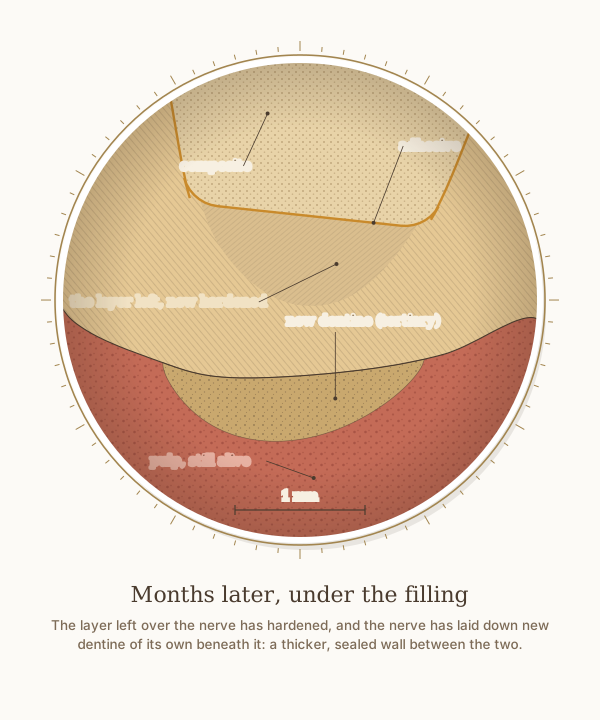
A close-up of the same measured molar months after the filling, with a 1 mm scale. Sealed under the composite, the layer of softened dentine left over the nerve has hardened, and the nerve has laid down new dentine of its own on the roof of the pulp chamber, so the wall between the filling and the nerve is thicker than on the day of treatment.
What the drawing is measured from
- In 32 teeth with deep decay, the decay was only partly removed, then the teeth were lined with calcium hydroxide and sealed for six to seven months. When they were reopened the dentine was dry, it was hard in 80 per cent of the 30 teeth still in the study, and the bacteria in it had decreased significantly. Maltz et al. 2002, Quintessence International. PubMed 1
- Under fillings placed after indirect pulp capping in children's back teeth, cone-beam CT measured about 0.5 mm of new dentine on the roof of the pulp chamber after six months, with no meaningful difference between calcium hydroxide, glass ionomer and MTA. Mathur et al. 2016, Indian Journal of Dental Research, randomised trial. DOI 2
- Tertiary dentine laid down by the surviving cells at the edge of the pulp in response to a stimulus such as decay is called reactionary dentine. Smith et al. 1995, International Journal of Developmental Biology, review. PubMed 3
- In deep lesions in teeth with healthy pulps, international consensus recommends selective removal to soft dentine: decayed dentine close to the pulp does not need to be removed, and the priorities are keeping healthy tooth tissue, achieving a seal and keeping the pulp healthy. Schwendicke et al. 2016, Advances in Dental Research, International Caries Consensus Collaboration. DOI 4
- The new dentine is drawn 0.5 mm thick at its thickest, the amount measured after six months in the trial above; the soft layer is drawn as it was left, now hardened.
Use this diagram
Free to use, including commercially, with credit.
This diagram is published under CC BY 4.0. You may reproduce it in articles, teaching material, presentations and patient information, print or online, without asking. The one condition is that you credit Dr. Khalid Aletaibi and link back to this page.
It is published this way on purpose. The measurement in this drawing is not something the people selling the treatment tend to publish, so it is more useful spread than kept.
Credit line
Months later, under the filling. Dr. Khalid Aletaibi, CC BY 4.0. https://khalidaletaibi.com/en/diagrams/composite-filling-later
HTML embed
<figure>
  <img src="https://khalidaletaibi.com/figures/composite-filling-later-en.svg" alt="Months later, under the filling" width="100%">
  <figcaption>
    Months later, under the filling. <a href="https://khalidaletaibi.com/en/diagrams/composite-filling-later">Dr. Khalid Aletaibi</a>,
    <a href="https://creativecommons.org/licenses/by/4.0/">CC BY 4.0</a>
  </figcaption>
</figure>