Measured diagram
Where should the edge of a crown sit against the gum?
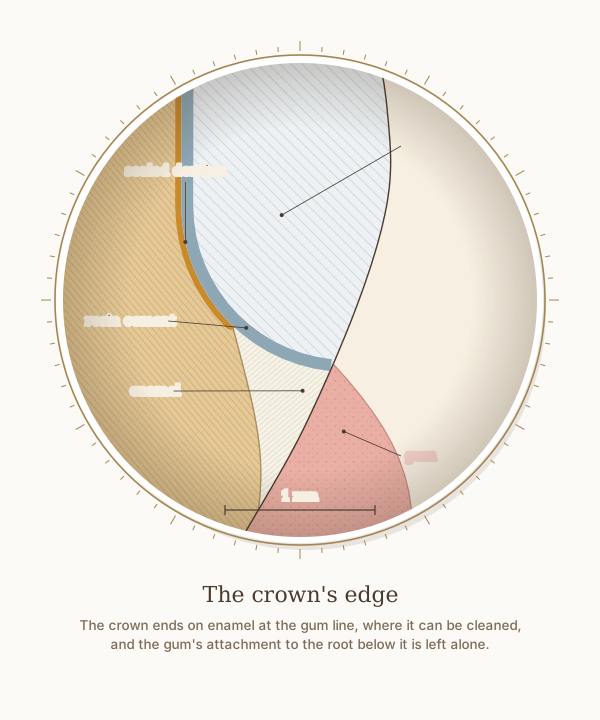
A close-up of the crown's tongue-side edge, from the same measured molar as the step-by-step figure, with a 1 mm scale. The crown ends on enamel at the gum line, where it can be cleaned and checked, and the gum's attachment to the root below it is left undisturbed.
What the drawing is measured from
- In 160 men followed for 26 years, filling margins placed below the gum line were detrimental to gum and periodontal health; the extra loss of attachment began slowly and could be detected 1 to 3 years after the restorations were placed. Schätzle et al. 2001, Journal of Clinical Periodontology. DOI
- The 2017 World Workshop replaced the term biologic width with supracrestal tissue attachment, meaning the junctional epithelium and the connective tissue above the bone, and found that restoration margins placed within that connective tissue attachment are associated with inflammation or loss of periodontal support. Jepsen et al. 2018, Journal of Periodontology, consensus report. DOI
- That attachment has no single width: a systematic review found means of 2.15 to 2.30 mm but a range from 0.2 to 6.73 mm between individuals. Schmidt et al. 2013, Journal of Clinical Periodontology. DOI
- The preparation follows the recommendations an evidence map of 73 articles consolidated for all-ceramic CAD/CAM back-tooth crowns: 1.5 to 2.0 mm of occlusal reduction, a chamfer of at least 1.0 mm, 1.0 to 1.5 mm of axial reduction, 12 degrees of total convergence and rounded internal angles. Deranek et al. 2025, Journal of Dental Education. DOI
- For glass-ceramics such as lithium disilicate, a systematic review of 83 laboratory studies found that etching with hydrofluoric acid followed by a silane is the most widely accepted surface treatment before a resin cement. Tian et al. 2014, Dental Materials. DOI
- Drawn as Dr. Khalid places the margin: at the gum line, on enamel, bonded with resin cement under rubber dam. The cement and the sealing are drawn 0.08 mm thick; both are representative rather than measured.
Use this diagram
Free to use, including commercially, with credit.
This diagram is published under CC BY 4.0. You may reproduce it in articles, teaching material, presentations and patient information, print or online, without asking. The one condition is that you credit Dr. Khalid Aletaibi and link back to this page.
It is published this way on purpose. The measurement in this drawing is not something the people selling the treatment tend to publish, so it is more useful spread than kept.
Credit line
The crown's edge. Dr. Khalid Aletaibi, CC BY 4.0. https://khalidaletaibi.com/en/diagrams/crown-margin
HTML embed
<figure>
  <img src="https://khalidaletaibi.com/figures/crown-margin-en.svg" alt="The crown's edge" width="100%">
  <figcaption>
    The crown's edge. <a href="https://khalidaletaibi.com/en/diagrams/crown-margin">Dr. Khalid Aletaibi</a>,
    <a href="https://creativecommons.org/licenses/by/4.0/">CC BY 4.0</a>
  </figcaption>
</figure>