Measured diagram
How is a crown placed on a root-filled tooth that has lost its walls?
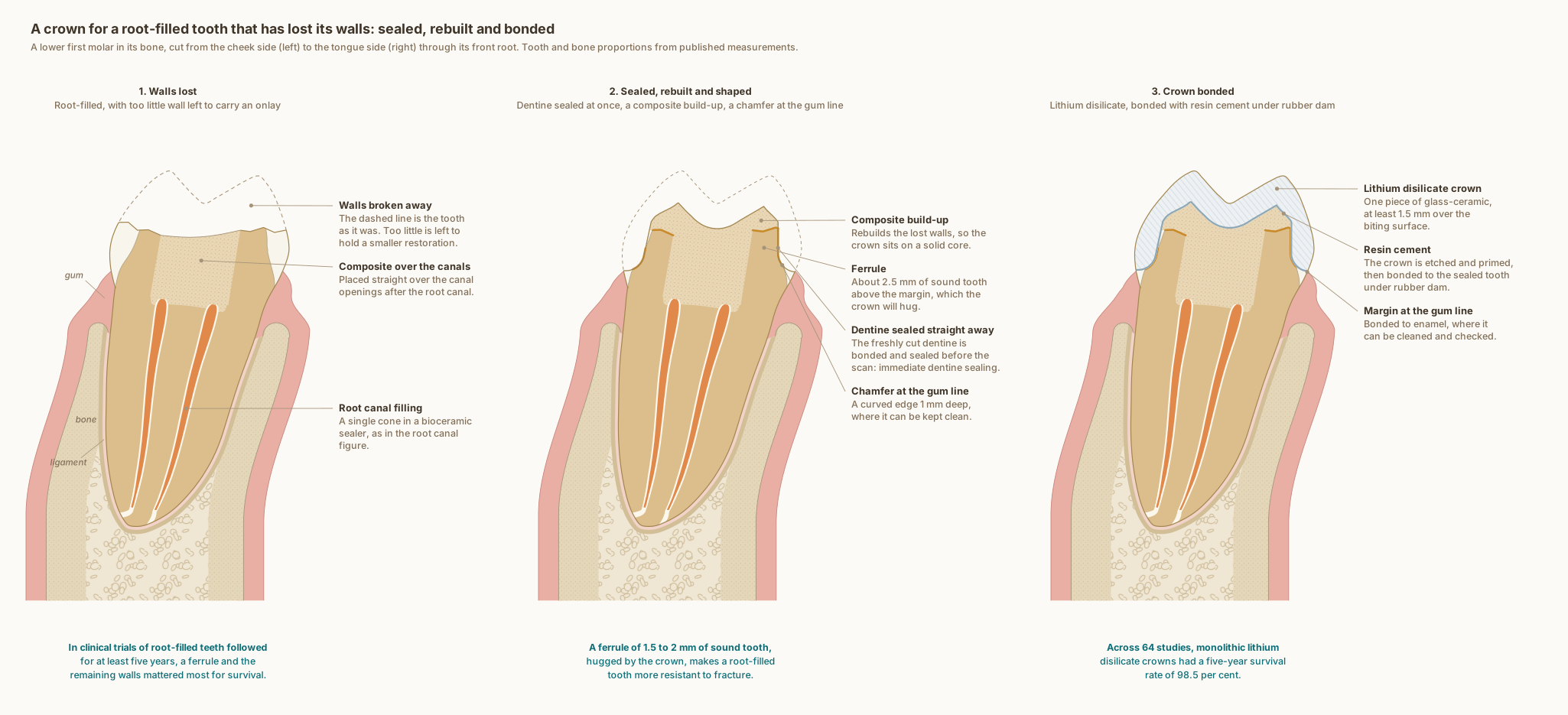
The same lower first molar in every panel, cut from the cheek side to the tongue side through its front root, with the gum, ligament and bone around it. The tooth and bone are drawn to published measurements; the broken walls, the build-up and the crown show what typically happens, not one patient's tooth.
What the drawing is measured from
- In a systematic review of 7 randomised trials and 1 prospective trial of root-filled teeth followed for at least five years, a ferrule and the remaining cavity walls were the main factors in the survival of the tooth and its restoration, and most studies found no benefit from a post. Naumann et al. 2018, Journal of Endodontics. DOI
- A literature review of laboratory, computer and clinical studies concluded that a ferrule of 1.5 to 2 mm improves the fracture resistance of root-filled teeth, that an incomplete ferrule is better than none, and that where no ferrule can be created at all a poor outcome is very likely. Juloski et al. 2012, Journal of Endodontics. DOI
- A systematic review and meta-analysis of 64 clinical studies, covering 8,051 all-ceramic and 3,509 metal-ceramic single crowns, estimated five-year survival at 98.5 per cent for monolithic lithium disilicate crowns, and found fewer ceramic fractures and chips with monolithic designs than with veneered ones. Pjetursson et al. 2026, The International Journal of Prosthodontics. DOI
- The preparation follows the recommendations an evidence map of 73 articles consolidated for all-ceramic CAD/CAM back-tooth crowns: 1.5 to 2.0 mm of occlusal reduction, a chamfer of at least 1.0 mm, 1.0 to 1.5 mm of axial reduction, 12 degrees of total convergence and rounded internal angles. Deranek et al. 2025, Journal of Dental Education. DOI
- Monolithic lithium disilicate crowns 2.0 mm thick withstood 17 times as many loading cycles in water as 1.0 mm crowns, and 0.5 mm crowns failed at the first; the authors suggest at least 1.5 mm for back-tooth crowns. Dhima et al. 2014, Journal of Prosthodontics, laboratory study. DOI
- Immediate dentine sealing means applying and curing a dentine bonding agent on the freshly cut dentine straight after the preparation, before the impression or scan; a review of more than 30 articles found that it appears to give better bond strength, fewer gaps, less bacterial leakage and less sensitivity. Magne 2005, Journal of Esthetic and Restorative Dentistry, review. DOI
- For glass-ceramics such as lithium disilicate, a systematic review of 83 laboratory studies found that etching with hydrofluoric acid followed by a silane is the most widely accepted surface treatment before a resin cement. Tian et al. 2014, Dental Materials. DOI
- In 160 men followed for 26 years, filling margins placed below the gum line were detrimental to gum and periodontal health; the extra loss of attachment began slowly and could be detected 1 to 3 years after the restorations were placed. Schätzle et al. 2001, Journal of Clinical Periodontology. DOI
- The case and the steps are drawn as Dr. Khalid works: a root-filled molar that has lost its walls, its canals filled as in the site's root canal figure (a single cone in a bioceramic sealer, with composite placed straight over the canal openings); immediate dentine sealing on the freshly cut dentine, then a composite build-up; a chamfer margin at the gum line; and a monolithic lithium disilicate crown bonded with resin cement under rubber dam. Where the walls broke is illustrative; it is set so that about 2.5 mm of sound tooth remains above the margin.
- The enamel follows one measured dataset for this tooth, from cone-beam CT of 45 lower first molars: 1.58 mm at the front cheek-side cusp tip and 0.98 mm at the central fossa, its thinnest point. Zhang et al. 2026, BMC Oral Health. DOI
- Two canals in the front root, as drawn, are found in 94.4 per cent of lower first molars, and three in 2.3 per cent, from a systematic review of 41 studies covering 18,781 teeth. de Pablo et al. 2010, Journal of Endodontics. DOI
- Bone level: on cone-beam CT of 100 lower molar sites in 25 adults, the crest of the bone sat 2.03 to 2.11 mm below the enamel edge of first molars, with 1.14 to 1.19 mm of bone over the cheek side of the root near its neck. Othman 2026, The Saudi Dental Journal. DOI
- The cheek-side cortical plate near the crest is drawn from cone-beam CT of lower first and second molars, where it measured 1.1 to 1.4 mm. Sayed et al. 2021, The Saudi Dental Journal. DOI
- Further from the crest, on cone-beam CT of 100 people, between the lower second premolar and first molar: the cheek-side plate measured 1.93 mm at 4 mm below the enamel edge and 2.36 mm at 6 mm, and the tongue-side plate 2.18 and 2.09 mm. Gupta et al. 2025, BMC Oral Health. DOI
- The gum is drawn 1.2 mm thick, within the 0.8 to 1.5 mm measured by ultrasound across lower teeth in 200 periodontally healthy men. Eger, Müller and Heinecke 1996, Journal of Clinical Periodontology. DOI
- The tooth follows the textbook descriptions of a lower first molar rather than a symmetrical outline: the crown leans toward the tongue on its root, the front root's tip lies under the front cheek-side cusp, the roof of the pulp chamber sits at the level of the enamel edge with the floor in the root trunk, and the ligament around the root is drawn 0.2 mm wide, under the quarter of a millimetre the textbooks give, inside a thin lining of compact bone. Wheeler's Dental Anatomy, Physiology and Occlusion, 11th edition, and Woelfel's Dental Anatomy.
- The roof of the pulp chamber sits at the level of the enamel edge, where it lay in 97 per cent of 100 lower molars measured on radiographs, and the drawing's distance from the cheek-side cusp tip to that roof, 6.36 mm, is the study's mean. Deutsch and Musikant 2004, Journal of Endodontics. DOI
Use this diagram
Free to use, including commercially, with credit.
This diagram is published under CC BY 4.0. You may reproduce it in articles, teaching material, presentations and patient information, print or online, without asking. The one condition is that you credit Dr. Khalid Aletaibi and link back to this page.
It is published this way on purpose. The measurement in this drawing is not something the people selling the treatment tend to publish, so it is more useful spread than kept.
Credit line
A crown for a root-filled tooth that has lost its walls: sealed, rebuilt and bonded. Dr. Khalid Aletaibi, CC BY 4.0. https://khalidaletaibi.com/en/diagrams/crown-steps
HTML embed
<figure>
  <img src="https://khalidaletaibi.com/figures/crown-steps-en.svg" alt="A crown for a root-filled tooth that has lost its walls: sealed, rebuilt and bonded" width="100%">
  <figcaption>
    A crown for a root-filled tooth that has lost its walls: sealed, rebuilt and bonded. <a href="https://khalidaletaibi.com/en/diagrams/crown-steps">Dr. Khalid Aletaibi</a>,
    <a href="https://creativecommons.org/licenses/by/4.0/">CC BY 4.0</a>
  </figcaption>
</figure>