Measured diagram
How thick is the enamel on an upper front tooth?
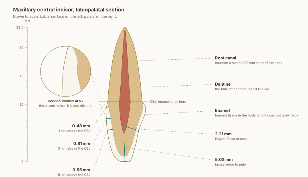
Drawn to scale from published measurements, with every labelled dimension measured back off the drawn geometry rather than asserted beside it. The enamel really is that thin at the neck.
What the drawing is measured from
- Labial enamel thickness at 1, 3 and 5 mm above the cementoenamel junction: 0.48, 0.81 and 0.95 mm. Cone-beam CT of 1,600 maxillary central incisors in patients aged 18 to 40, Diagnostics 2024, PMC11592583. In agreement with micro-CT of 20 central incisors, which found a range of 0.461 to 0.966 mm. Enamel also thins significantly with age, so these are not lifetime values. PubMed
- Pulp distances measured on cone-beam CT across 200 teeth: palatal fossa to the pulp chamber 2.21 mm, incisal edge to the pulp 5.02 mm. Kohok et al. 2025, PMID 41193532. PubMed
- In anterior teeth the major apical foramen sits a mean 0.39 mm short of the anatomic apex and coincides with the tip in only about 40 per cent of teeth, from 1,331 roots. Martos et al. 2009, PMID 19220517. Cementum is a median 97 micrometres cervically and 217 micrometres apically, PMID 34999579, so it is drawn as the root outline rather than as its own layer. Crown and root proportions follow Wheeler and Nelson averages. PubMed
- Two things on this drawing are anatomically representative but are not taken from published measurements, and are therefore not labelled with a number: the palatal enamel profile, and the width of the pulp space. Only the labelled values are sourced.
Use this diagram
Free to use, including commercially, with credit.
This diagram is published under CC BY 4.0. You may reproduce it in articles, teaching material, presentations and patient information, print or online, without asking. The one condition is that you credit Dr. Khalid Aletaibi and link back to this page.
It is published this way on purpose. The measurement in this drawing is not something the people selling the treatment tend to publish, so it is more useful spread than kept.
Credit line
Maxillary central incisor, labiopalatal section. Dr. Khalid Aletaibi, CC BY 4.0. https://khalidaletaibi.com/en/diagrams/incisor-anatomy
HTML embed
<figure>
  <img src="https://khalidaletaibi.com/figures/incisor-anatomy-en.svg" alt="Maxillary central incisor, labiopalatal section" width="100%">
  <figcaption>
    Maxillary central incisor, labiopalatal section. <a href="https://khalidaletaibi.com/en/diagrams/incisor-anatomy">Dr. Khalid Aletaibi</a>,
    <a href="https://creativecommons.org/licenses/by/4.0/">CC BY 4.0</a>
  </figcaption>
</figure>