Measured diagram
What does a lower first molar look like in cross section?
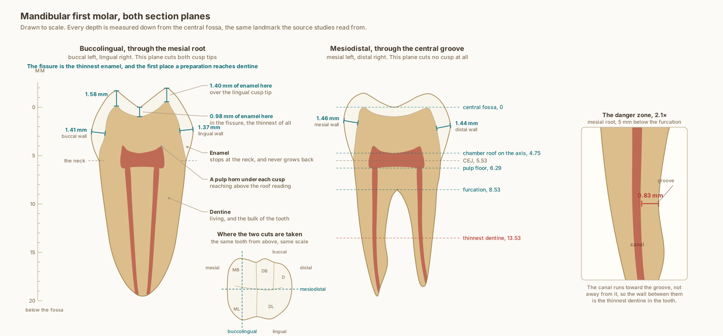
Drawn to scale, with every depth measured down from the central fossa, which is the landmark the source studies read from. Each stated dimension is measured back off the drawn geometry rather than asserted beside it.
What the drawing is measured from
- Enamel comes from one complete landmark dataset for this tooth, so that no value is invented and none is stitched together from a study that measured something else. Cone-beam CT at 0.16 mm voxel, 45 mandibular first permanent molars: mesiobuccal cusp tip 1.58 mm and its buccal surface 1.44 mm, mesiolingual cusp tip 1.40 mm and its lingual surface 1.34 mm, both marginal ridges 1.48 mm, central fossa 0.98 mm. Zhang et al. 2026, BMC Oral Health. DOI
- That sample is unworn first molars in children whose crowns had formed but were not yet in occlusion, so the figures are maximum values before any wear. Sources genuinely disagree here and it is worth saying so: micro-CT at 28 micrometres on unworn adult teeth reads the same two cusp tips at 0.94 and 1.12 mm, and histology on 25 first molars reads 1.16 and 1.13 with the fossa at 0.87. All three agree the central fossa is the thinnest point, and that it is the first place a preparation reaches dentine. PubMed
- Pulp landmarks measured on cone-beam CT across 104 mandibular molars, read to 0.05 mm: central fossa to chamber roof 4.75 mm, chamber height 1.53 mm, floor 6.29 mm, furcation 8.53 mm. Azim et al. 2014, PMID 25146010. PubMed
- The thinnest dentine in the mesial root sits 5 mm below the furcation in over 85 per cent of teeth, with a distal wall of 0.83 mm. Cone-beam CT, 624 teeth, PMC11208803. The apical foramen sits a mean 0.6 mm from the anatomical apex and deviates from it in 85 per cent of teeth, from 238 extracted teeth, PMID 40634457. PubMed
- Crown and root proportions follow Wheeler and Nelson averages. Cusp height above the fossa is not separately published; it is solved at 1.97 mm, the only value at which the crown height, the furcation depth and the root trunk measurements are mutually consistent.
Use this diagram
Free to use, including commercially, with credit.
This diagram is published under CC BY 4.0. You may reproduce it in articles, teaching material, presentations and patient information, print or online, without asking. The one condition is that you credit Dr. Khalid Aletaibi and link back to this page.
It is published this way on purpose. The measurement in this drawing is not something the people selling the treatment tend to publish, so it is more useful spread than kept.
Credit line
Mandibular first molar, both section planes. Dr. Khalid Aletaibi, CC BY 4.0. https://khalidaletaibi.com/en/diagrams/molar-anatomy
HTML embed
<figure>
  <img src="https://khalidaletaibi.com/figures/molar-anatomy-en.svg" alt="Mandibular first molar, both section planes" width="100%">
  <figcaption>
    Mandibular first molar, both section planes. <a href="https://khalidaletaibi.com/en/diagrams/molar-anatomy">Dr. Khalid Aletaibi</a>,
    <a href="https://creativecommons.org/licenses/by/4.0/">CC BY 4.0</a>
  </figcaption>
</figure>