Measured diagram
What is inside a gum pocket?
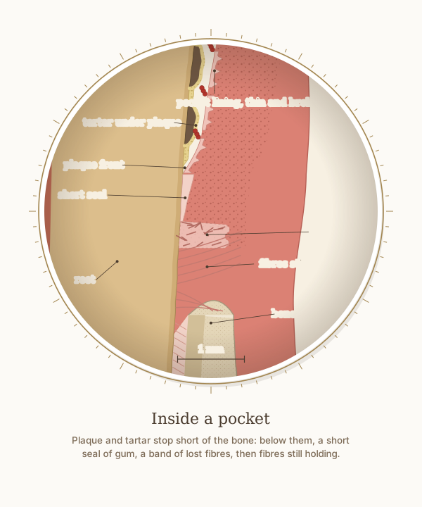
A close-up of the tongue side of the 5 mm pocket, with a 1 mm scale. Plaque and tartar stop short of the bone: below them lie a short seal of gum, a band of lost fibres, then fibres still holding the tooth.
What the drawing is measured from
- Plaque never reaches the bone: on 48 extracted teeth the front of the plaque lay 0.2 to 2.0 mm from the remaining attachment fibres, and in autopsy material never less than 0.5 mm or more than 2.7 mm from the bone. DOI 1
- Inside a pocket, between the last epithelium and the first intact fibres there was a zone of destroyed fibres averaging 0.43 mm wide, in almost every tooth examined. DOI 2
- The pocket lining does not attach to the tooth: it is thin, ridged and full of inflammatory cells, with breaks in places (a study of experimental pockets in dogs). DOI 3
- Tartar always carries plaque: a living film of plaque covers mineralised tartar deposits, above and below the gum. DOI 4
- Two kinds of tartar: above the gum it is yellow-white, layered and about 37% mineral by volume; below the gum it is dark brown, uniform and about 58% mineral, usually a thin, smooth veneer on the root (Friskopp and Isacsson 1984; Woelfel; Gurgan and Bilgin 2005). DOI 5
- Where the probe stops in a pocket: in untreated pockets the tip went 0.29 mm (Magnusson and Listgarten 1980) and 0.45 mm (Fowler 1982) past the epithelium into inflamed tissue, well short of the bone; after treatment it stopped inside the epithelium. DOI 6
- The tooth: a lower central incisor in section, crown 9.5 mm and root 12.5 mm (Wheeler's Dental Anatomy, Table 7.1), with the lip side on the left. Pockets and openings are drawn slightly open so they can be seen; no number on the drawing is a measurement of one person.
Use this diagram
Free to use, including commercially, with credit.
This diagram is published under CC BY 4.0. You may reproduce it in articles, teaching material, presentations and patient information, print or online, without asking. The one condition is that you credit Dr. Khalid Aletaibi and link back to this page.
It is published this way on purpose. The measurement in this drawing is not something the people selling the treatment tend to publish, so it is more useful spread than kept.
Credit line
Inside a pocket. Dr. Khalid Aletaibi, CC BY 4.0. https://khalidaletaibi.com/en/diagrams/periodontal-pocket
HTML embed
<figure>
  <img src="https://khalidaletaibi.com/figures/periodontal-pocket-en.svg" alt="Inside a pocket" width="100%">
  <figcaption>
    Inside a pocket. <a href="https://khalidaletaibi.com/en/diagrams/periodontal-pocket">Dr. Khalid Aletaibi</a>,
    <a href="https://creativecommons.org/licenses/by/4.0/">CC BY 4.0</a>
  </figcaption>
</figure>