Measured diagram
What do the numbers on a gum chart mean?
The reading is the distance from the gum's edge to where the probe stops, shown by the bracket beside each probe. Healthy gums read 1 to 3 mm and do not bleed; the aim of treatment is no pocket deeper than 4 mm that bleeds and none of 6 mm or more.
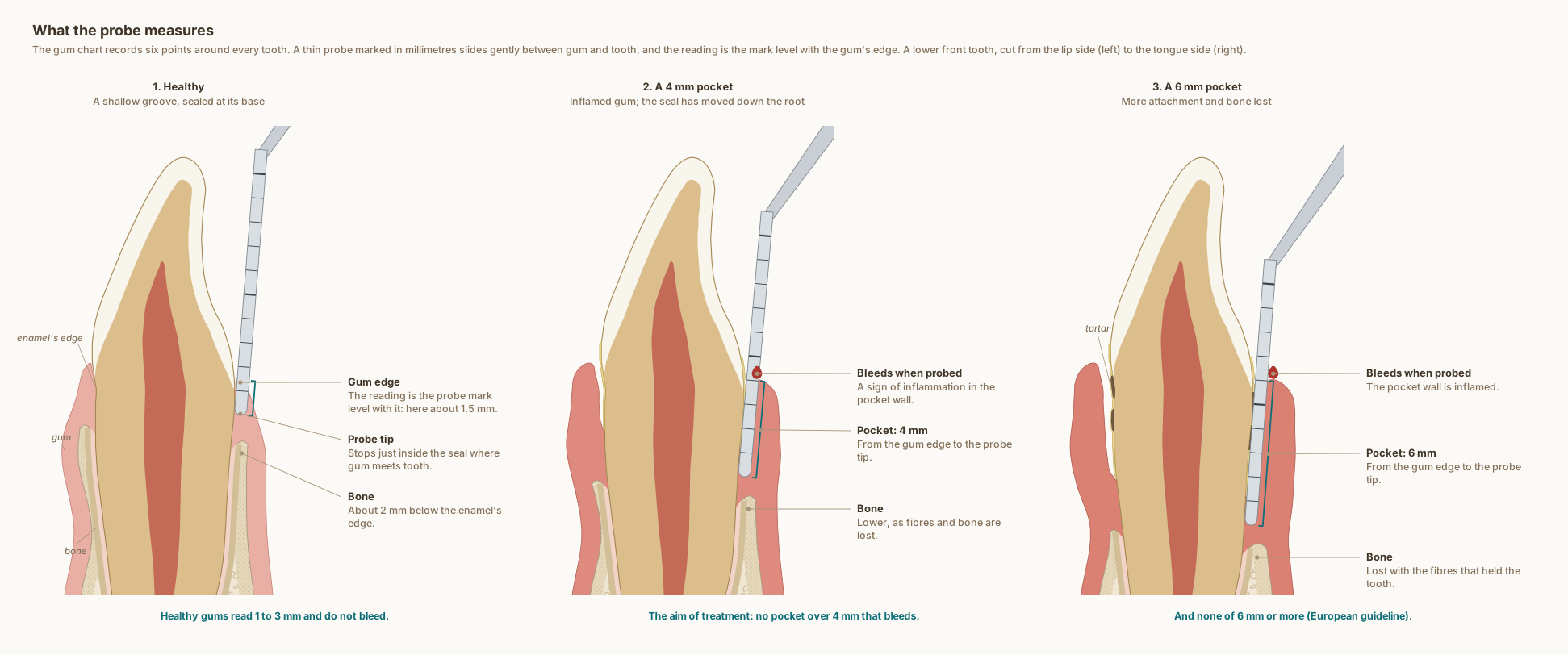
What the drawing is measured from
- Healthy gum anatomy: the gum edge sits about 1 mm above the enamel's edge and healthy gums probe 1 to 3 mm (Woelfel's Dental Anatomy). Under a groove about 0.7 mm deep lie about 1 mm of attaching epithelium and 1 mm of fibres above the bone, Gargiulo's autopsy means, which vary from person to person. DOI 1
- Gum thickness: at the lower front teeth the gum averaged 0.71 mm thick, and healthy pockets read 1.79 mm on average, in 187 people with healthy gums. DOI 2
- The probe: commercial probe tips measured 0.28 to 0.70 mm across, and a light force of about 0.25 N is used; at higher forces healthy gums bleed more often. DOI 3
- Where the probe stops in health: at 22 sites on front teeth, the probe tip stopped inside the junctional epithelium, on average 0.25 mm short of its end. DOI 4
- Where the probe stops in a pocket: in untreated pockets the tip went 0.29 mm (Magnusson and Listgarten 1980) and 0.45 mm (Fowler 1982) past the epithelium into inflamed tissue, well short of the bone; after treatment it stopped inside the epithelium. DOI 5
- Bone around lower central incisors: in CBCT scans of 50 women without gum disease, the bone crest lay about 2.2 to 2.8 mm below the enamel's edge, and the bone on the lip side was thin, about 0.5 to 0.75 mm including the ligament, along most of the root. DOI 6
- The European guideline (EFP S3): the aim is no pocket deeper than 4 mm that bleeds and none of 6 mm or more; sites are re-assessed after healing; hand or powered instruments, or both; supportive care every 3 to at most 12 months. DOI 7
- The tooth: a lower central incisor in section, crown 9.5 mm and root 12.5 mm (Wheeler's Dental Anatomy, Table 7.1), with the lip side on the left. Pockets and openings are drawn slightly open so they can be seen; no number on the drawing is a measurement of one person.
Use this diagram
Free to use, including commercially, with credit.
This diagram is published under CC BY 4.0. You may reproduce it in articles, teaching material, presentations and patient information, print or online, without asking. The one condition is that you credit Dr. Khalid Aletaibi and link back to this page.
It is published this way on purpose. The measurement in this drawing is not something the people selling the treatment tend to publish, so it is more useful spread than kept.
Credit line
What the probe measures. Dr. Khalid Aletaibi, CC BY 4.0. https://khalidaletaibi.com/en/diagrams/periodontal-probe-chart
HTML embed
<figure>
  <img src="https://khalidaletaibi.com/figures/periodontal-probe-chart-en.svg" alt="What the probe measures" width="100%">
  <figcaption>
    What the probe measures. <a href="https://khalidaletaibi.com/en/diagrams/periodontal-probe-chart">Dr. Khalid Aletaibi</a>,
    <a href="https://creativecommons.org/licenses/by/4.0/">CC BY 4.0</a>
  </figcaption>
</figure>