Measured diagram
What is the difference between an inlay, an onlay, an overlay and a crown?
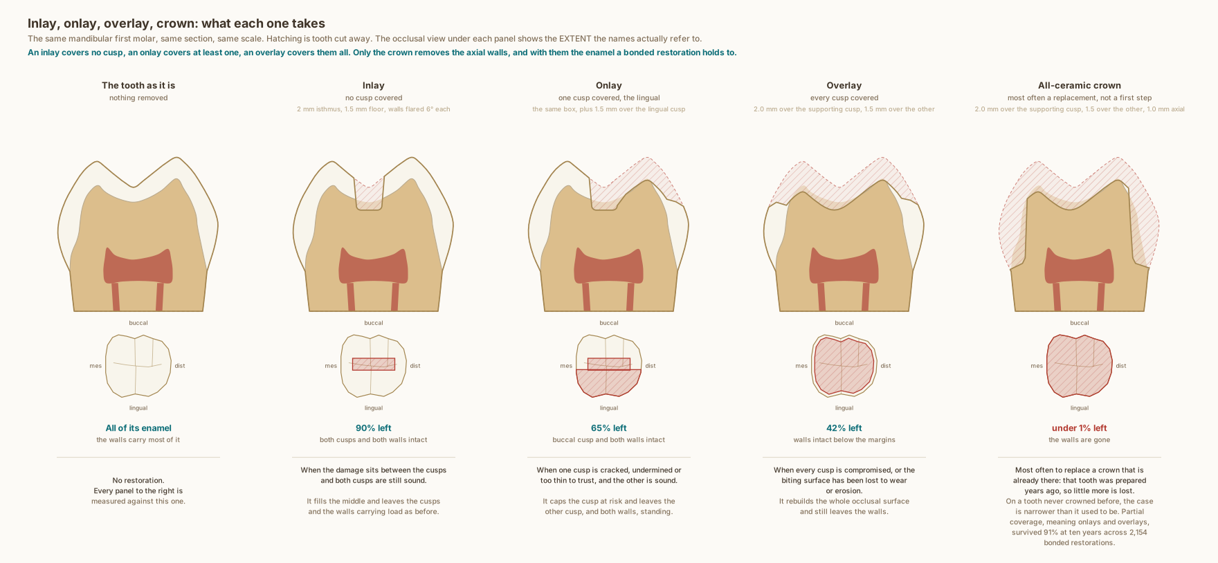
The same mandibular first molar in every panel, drawn to scale from measured anatomy. The percentages are areas measured off these drawings by scanline every 0.005 millimetres, within this one section, and are not whole-tooth volumes.
What the drawing is measured from
- Preparation dimensions follow concordant published guidelines: pulpal floor 1.5 to 2.0 mm, isthmus 2.0 mm, a 90 degree butt-joint cavosurface with no bevel, rounded internal line angles, and walls flared 10 to 12 degrees in total. Cuspal reduction 1.5 to 2.0 mm, drawn here at the shallow end of that range.
- Covering a cusp is advised below roughly 2 mm of remaining thickness at the base of a supporting cusp and 3 mm at a non-supporting one. In a lower molar the buccal cusps support and work in compression, while the lingual cusps work in tension and are the ones that fail, which is why the onlay here covers the lingual cusp.
- Those coverage thresholds date from the era of metal restorations and are being revisited. Bonded lithium disilicate onlays left on thin cusps did not fracture under five clinical years of chewing simulation, and the authors argue for preparations that keep more tooth. Griffis et al. 2020, Journal of Esthetic and Restorative Dentistry. DOI
- Bonded ceramic inlays, onlays and overlays survived 92 to 95 per cent at five years across 5,811 restorations, and 91 per cent at ten years across 2,154, with fracture the commonest failure and endodontic complications the next. Systematic review and meta-analysis, Morimoto et al. 2016, Journal of Dental Research. DOI
- Whole-tooth volumes, for comparison with the section areas on the drawing: adhesive and inlay preparations removed 5.5 to 27.2 per cent of the coronal tooth and complete crowns 67.5 to 75.6 per cent. Measured gravimetrically on resin replica teeth, 10 preparations per design, not on natural teeth. Edelhoff and Sorensen 2002, PMID 12186346. PubMed
- Enamel thickness at named occlusal landmarks, from cone-beam CT at 0.16 mm voxel across 45 mandibular first permanent molars: mesiobuccal cusp tip 1.58 mm, mesiolingual 1.40 mm, both marginal ridges 1.48 mm, and the central fossa 0.98 mm, the thinnest landmark measured. Zhang et al. 2026, BMC Oral Health. DOI
- Which restoration suits a given tooth is a clinical judgement made case by case, on the tooth in front of you.
Use this diagram
Free to use, including commercially, with credit.
This diagram is published under CC BY 4.0. You may reproduce it in articles, teaching material, presentations and patient information, print or online, without asking. The one condition is that you credit Dr. Khalid Aletaibi and link back to this page.
It is published this way on purpose. The measurement in this drawing is not something the people selling the treatment tend to publish, so it is more useful spread than kept.
Credit line
Inlay, onlay, overlay, crown: what each one takes. Dr. Khalid Aletaibi, CC BY 4.0. https://khalidaletaibi.com/en/diagrams/restoration-ladder
HTML embed
<figure>
  <img src="https://khalidaletaibi.com/figures/restoration-ladder-en.svg" alt="Inlay, onlay, overlay, crown: what each one takes" width="100%">
  <figcaption>
    Inlay, onlay, overlay, crown: what each one takes. <a href="https://khalidaletaibi.com/en/diagrams/restoration-ladder">Dr. Khalid Aletaibi</a>,
    <a href="https://creativecommons.org/licenses/by/4.0/">CC BY 4.0</a>
  </figcaption>
</figure>