Measured diagram
Where does an infected root canal meet the bone?
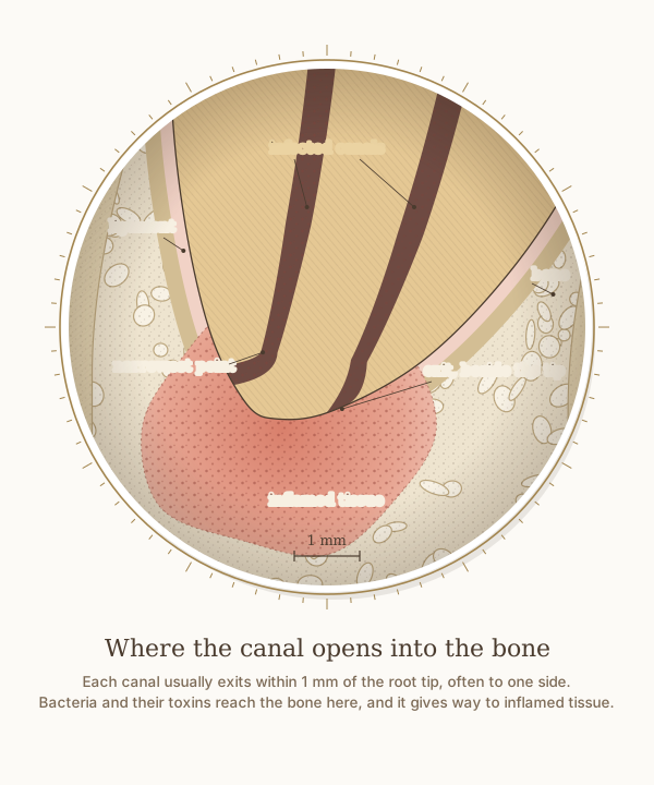
A close-up of the front root's tip from the same measured molar as the step-by-step figure, with a 1 mm scale. Each canal narrows just before it exits, usually a little to one side of the tip, and that exit is where bacteria and their toxins reach the bone.
What the drawing is measured from
- Where the canal exits: across 1,331 roots, the main exit of the canal lay a mean 0.80 mm from the root tip in lower molars, always within 1 mm, and sat exactly at the tip in only 40 per cent of teeth; when it lay to one side, the cheek side was the commonest. Martos et al. 2009, International Endodontic Journal. DOI
- The narrowest point of the canal: in micro-CT scans of 56 extracted molars, in canals with a single narrowing, it lay a mean 0.76 mm inside the canal's exit. Hu et al. 2026, BMC Oral Health. DOI
- The inflammation at the root tip comes from inside the tooth: apical periodontitis is a chronic inflammation of the tissues around the root tip caused by agents of endodontic origin, above all infection inside the root canal. Nair 2006, International Endodontic Journal, review. DOI
- The ligament that holds the root in its socket is drawn 0.2 mm wide, under the quarter of a millimetre the textbook gives, and the socket is lined by a thin layer of compact bone, the lamina dura, which shows on an X-ray as a white line around the root. Woelfel's Dental Anatomy.
- The size and outline of the inflamed area are representative rather than measured from a study, so they carry no number. On an X-ray it shows as a dark area around the root tip, because the inflamed tissue has replaced bone.
Use this diagram
Free to use, including commercially, with credit.
This diagram is published under CC BY 4.0. You may reproduce it in articles, teaching material, presentations and patient information, print or online, without asking. The one condition is that you credit Dr. Khalid Aletaibi and link back to this page.
It is published this way on purpose. The measurement in this drawing is not something the people selling the treatment tend to publish, so it is more useful spread than kept.
Credit line
Where the canal opens into the bone. Dr. Khalid Aletaibi, CC BY 4.0. https://khalidaletaibi.com/en/diagrams/root-canal-root-tip
HTML embed
<figure>
  <img src="https://khalidaletaibi.com/figures/root-canal-root-tip-en.svg" alt="Where the canal opens into the bone" width="100%">
  <figcaption>
    Where the canal opens into the bone. <a href="https://khalidaletaibi.com/en/diagrams/root-canal-root-tip">Dr. Khalid Aletaibi</a>,
    <a href="https://creativecommons.org/licenses/by/4.0/">CC BY 4.0</a>
  </figcaption>
</figure>