Measured diagram
What happens to the tooth and the bone during root canal treatment?
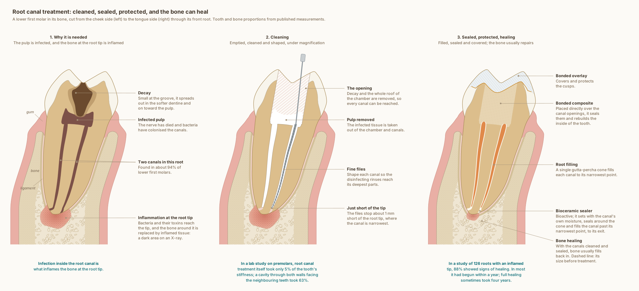
The same lower first molar in every panel, cut from the cheek side to the tongue side through its front root, with the gum, ligament and bone around it. The tooth and bone are drawn to published measurements; the three stages show what typically happens, not one patient's tooth.
What the drawing is measured from
- Two canals in the front root, as drawn, are found in 94.4 per cent of lower first molars, and three in 2.3 per cent, from a systematic review of 41 studies covering 18,781 teeth. de Pablo et al. 2010, Journal of Endodontics. DOI
- The inflammation at the root tip comes from inside the tooth: apical periodontitis is a chronic inflammation of the tissues around the root tip caused by agents of endodontic origin, above all infection inside the root canal. Nair 2006, International Endodontic Journal, review. DOI
- The note under the second panel comes from a laboratory study of 42 extracted upper second premolars, loaded without breaking them: the root canal procedures themselves reduced the tooth's relative stiffness by 5 per cent, an occlusal cavity by 20 per cent, and a cavity through both walls facing the neighbouring teeth by 63 per cent. Reeh, Messer and Douglas 1989, Journal of Endodontics. DOI
- The note under the third panel: of 126 roots treated with an inflamed tip and followed yearly for up to four years, 111 (88 per cent) showed signs of healing. Healing had begun within the first year in at least 89 per cent of those, and complete healing sometimes took four years. Ørstavik 1996, International Endodontic Journal. DOI
- Where the canal exits: across 1,331 roots, the main exit of the canal lay a mean 0.80 mm from the root tip in lower molars, always within 1 mm, and sat exactly at the tip in only 40 per cent of teeth; when it lay to one side, the cheek side was the commonest. Martos et al. 2009, International Endodontic Journal. DOI
- The narrowest point of the canal: in micro-CT scans of 56 extracted molars, in canals with a single narrowing, it lay a mean 0.76 mm inside the canal's exit. Hu et al. 2026, BMC Oral Health. DOI
- The root filling is drawn as Dr. Khalid places it: a single gutta-percha cone that ends at the canal's narrowest point, in a bioceramic sealer that fills the canal beyond it to the exit. In 307 teeth filled this way in a private practice and followed for an average of 30 months, overall success was 90.9 per cent; sealer passed beyond the root tip in 47.4 per cent of them, with no significant effect on the outcome. Chybowski et al. 2018, Journal of Endodontics, retrospective study. DOI
- Bioceramic (hydraulic calcium silicate) sealers were compared with other sealer types across 55 clinical reports covering 5,913 teeth: success rates were comparable, with no increase in pain after treatment. Donnermeyer et al. 2026, Journal of Dentistry, systematic review and meta-analysis. DOI
- In dogs' teeth with induced lesions at the root tip, a bioceramic sealer pushed beyond the tip on purpose produced less inflammation and more new bone at four months than a resin-based calcium silicate sealer. Elsayed et al. 2025, Clinical Oral Investigations, animal study. DOI
- Bone level: on cone-beam CT of 100 lower molar sites in 25 adults, the crest of the bone sat 2.03 to 2.11 mm below the enamel edge of first molars, with 1.14 to 1.19 mm of bone over the cheek side of the root near its neck. Othman 2026, The Saudi Dental Journal. DOI
- Bone beyond the root tip: on high-resolution cone-beam CT of 1,400 teeth, the lower first molar had 4.45 mm of bone from the exit of the cheek-side front canal to the cheek-side cortical plate, and 5.63 mm from the exit of the tongue-side front canal to the tongue-side plate. Porto et al. 2020, Journal of Applied Oral Science. DOI
- The cheek-side cortical plate near the crest is drawn from cone-beam CT of lower first and second molars, where it measured 1.1 to 1.4 mm. Sayed et al. 2021, The Saudi Dental Journal. DOI
- Further from the crest, on cone-beam CT of 100 people, between the lower second premolar and first molar: the cheek-side plate measured 1.93 mm at 4 mm below the enamel edge and 2.36 mm at 6 mm, and the tongue-side plate 2.18 and 2.09 mm. Gupta et al. 2025, BMC Oral Health. DOI
- The gum is drawn 1.2 mm thick, within the 0.8 to 1.5 mm measured by ultrasound across lower teeth in 200 periodontally healthy men. Eger, Müller and Heinecke 1996, Journal of Clinical Periodontology. DOI
- The tooth follows the textbook descriptions of a lower first molar rather than a symmetrical outline: the crown leans toward the tongue on its root, the front root's tip lies under the front cheek-side cusp, the roof of the pulp chamber sits at the level of the enamel edge with the floor in the root trunk, and the ligament around the root is drawn 0.2 mm wide, under the quarter of a millimetre the textbooks give, inside a thin lining of compact bone. Wheeler's Dental Anatomy, Physiology and Occlusion, 11th edition, and Woelfel's Dental Anatomy.
- Two choices in the drawing are clinical rather than measured, and both are drawn as Dr. Khalid works. The opening removes the whole roof of the pulp chamber, leaving no ledge over the pulp horns, so every canal entrance lies in view; its walls flare slightly and stop short of the cusp tips. The tooth is then rebuilt with bonded composite placed directly over the canal entrances, filling the chamber and the opening, and a bonded overlay covering the cusps.
- The roof of the pulp chamber sits at the level of the enamel edge, where it lay in 97 per cent of 100 lower molars measured on radiographs, and the drawing's distance from the cheek-side cusp tip to that roof, 6.36 mm, is the study's mean. Deutsch and Musikant 2004, Journal of Endodontics. DOI
Use this diagram
Free to use, including commercially, with credit.
This diagram is published under CC BY 4.0. You may reproduce it in articles, teaching material, presentations and patient information, print or online, without asking. The one condition is that you credit Dr. Khalid Aletaibi and link back to this page.
It is published this way on purpose. The measurement in this drawing is not something the people selling the treatment tend to publish, so it is more useful spread than kept.
Credit line
Root canal treatment: cleaned, sealed, protected, and the bone can heal. Dr. Khalid Aletaibi, CC BY 4.0. https://khalidaletaibi.com/en/diagrams/root-canal-steps
HTML embed
<figure>
  <img src="https://khalidaletaibi.com/figures/root-canal-steps-en.svg" alt="Root canal treatment: cleaned, sealed, protected, and the bone can heal" width="100%">
  <figcaption>
    Root canal treatment: cleaned, sealed, protected, and the bone can heal. <a href="https://khalidaletaibi.com/en/diagrams/root-canal-steps">Dr. Khalid Aletaibi</a>,
    <a href="https://creativecommons.org/licenses/by/4.0/">CC BY 4.0</a>
  </figcaption>
</figure>