Measured diagram
Where does a porcelain veneer end at the gum, and why does it stay on enamel?
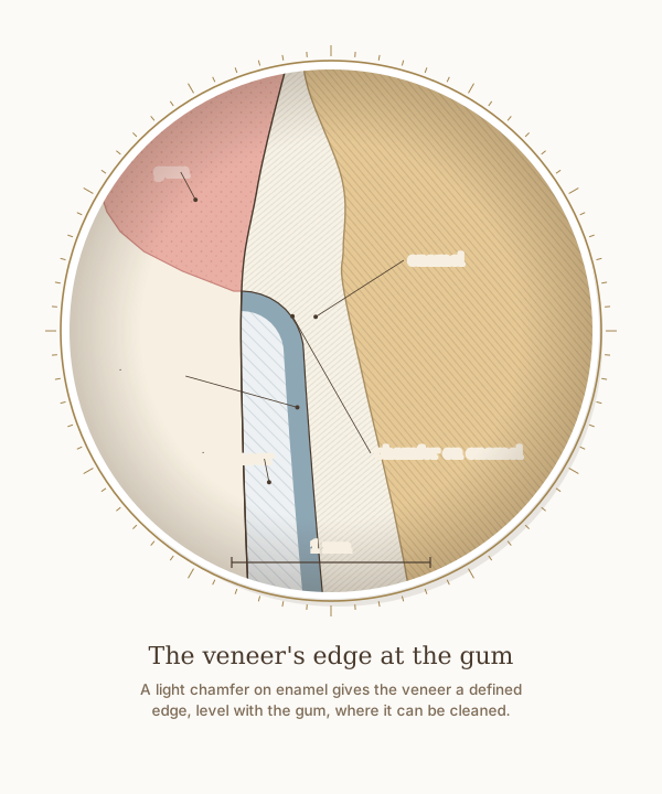
A close-up of the gum-line edge of the feldspathic veneer in the step-by-step figure, with a 1 mm scale. The light chamfer is cut on enamel and ends level with the gum, where the edge can be cleaned and the gum's attachment below it is left alone; the porcelain is bonded with resin cement.
What the drawing is measured from
- The preparation follows the moderate approach: reduction guided by the mock-up, a light chamfer of 0.3 to 0.4 mm at the margin, about 0.4 mm near the gum and 0.6 mm toward the edge for refractory-die porcelain, 1.0 to 1.5 mm of incisal clearance and a palatal butt margin. Where the mock-up adds volume, most of the tooth surface is left untouched, and a chamfer cut about 0.5 mm from a retraction cord ends level with the gum. Magne and Belser 2022, Biomimetic Restorative Dentistry, volume 2, pages 624 to 636.
- At the upper central incisors of 88 periodontally healthy adults, the gum overlapped the enamel by 1.41 mm, the bone crest lay 2.05 mm from the cementoenamel junction, the free gingiva was 1.12 mm thick at its base and the bone 0.45 mm thick at the crest. Alpiste-Illueca 2004, The International Journal of Periodontics and Restorative Dentistry. PubMed
- Labial enamel thickness at 1, 3 and 5 mm above the cementoenamel junction: 0.48, 0.81 and 0.95 mm. Cone-beam CT of 1,600 maxillary central incisors in patients aged 18 to 40, Diagnostics 2024, PMC11592583. In agreement with micro-CT of 20 central incisors, which found a range of 0.461 to 0.966 mm. Enamel also thins significantly with age, so these are not lifetime values. DOI
- Measured by electron microscopy on 40 upper central incisors from patients aged 30 to 69, the enamel was 0.31, 0.54 and 0.75 mm thick at 1, 3 and 5 mm above the cementoenamel junction, thinner than the CT values the drawing uses and thinning further from about age 50; near the gum line, a 0.3 mm chamfer can therefore come close to dentine. Atsu et al. 2005, The Journal of Prosthetic Dentistry. DOI
- In 62 lithium disilicate veneers in 12 patients, measured on silicone replicas by micro-CT, the median gap at the margin was 100 micrometres without preparation and 140 micrometres with minimal preparation, and the internal gap averaged about 0.17 to 0.22 mm. Karagözoğlu, Toksavul and Toman 2016, Quintessence International. DOI
- In 160 men followed for 26 years, filling margins placed below the gum line were detrimental to gum and periodontal health; the extra loss of attachment began slowly and could be detected 1 to 3 years after the restorations were placed. Schätzle et al. 2001, Journal of Clinical Periodontology. DOI
- The 2017 World Workshop replaced the term biologic width with supracrestal tissue attachment, meaning the junctional epithelium and the connective tissue above the bone, and found that restoration margins placed within that connective tissue attachment are associated with inflammation or loss of periodontal support. Jepsen et al. 2018, Journal of Periodontology, consensus report. DOI
- Drawn as Dr. Khalid places a veneer's edge: a light chamfer finish margin is always cut, here 0.3 mm deep for feldspathic porcelain, placed at the gum line on enamel. The cement is drawn 0.1 mm thick, close to the gaps measured at veneer margins; it is representative, not measured on this tooth.
Use this diagram
Free to use, including commercially, with credit.
This diagram is published under CC BY 4.0. You may reproduce it in articles, teaching material, presentations and patient information, print or online, without asking. The one condition is that you credit Dr. Khalid Aletaibi and link back to this page.
It is published this way on purpose. The measurement in this drawing is not something the people selling the treatment tend to publish, so it is more useful spread than kept.
Credit line
The veneer's edge at the gum. Dr. Khalid Aletaibi, CC BY 4.0. https://khalidaletaibi.com/en/diagrams/veneer-margin
HTML embed
<figure>
  <img src="https://khalidaletaibi.com/figures/veneer-margin-en.svg" alt="The veneer's edge at the gum" width="100%">
  <figcaption>
    The veneer's edge at the gum. <a href="https://khalidaletaibi.com/en/diagrams/veneer-margin">Dr. Khalid Aletaibi</a>,
    <a href="https://creativecommons.org/licenses/by/4.0/">CC BY 4.0</a>
  </figcaption>
</figure>