Measured diagram
How much enamel does a veneer remove, and does the porcelain matter?
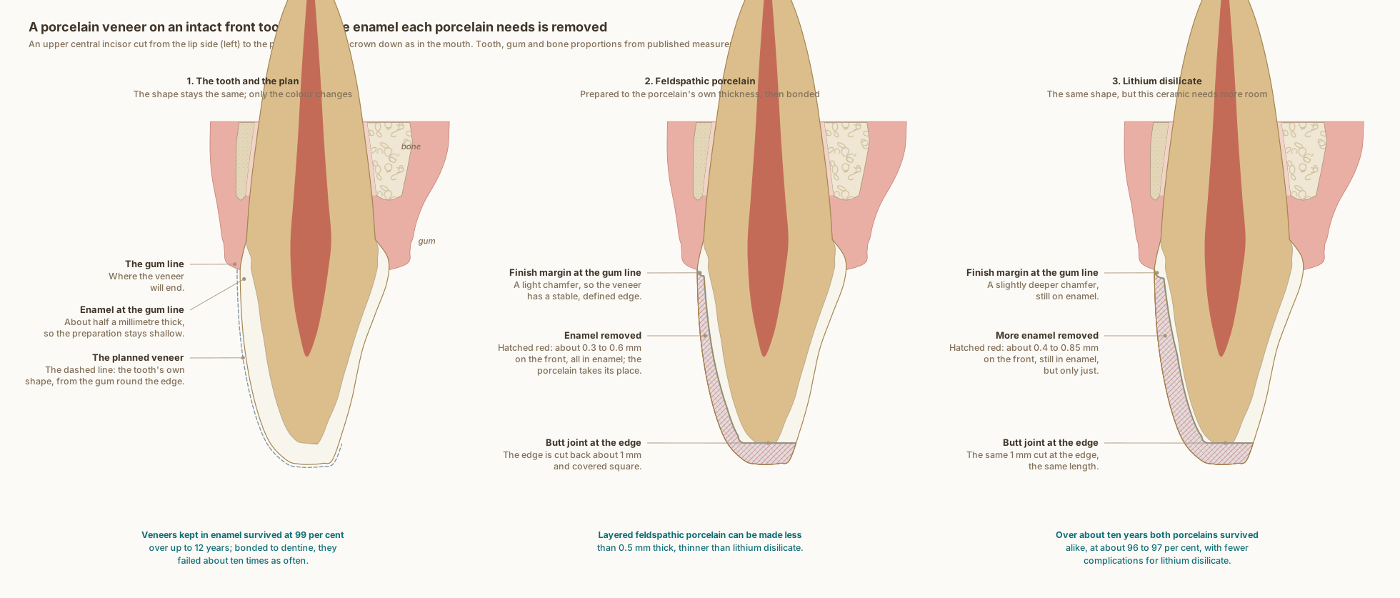
The same upper central incisor in every panel, cut from the lip side to the palate side, with the gum, ligament and bone around it. The tooth and the tissues at its neck are drawn to published measurements; the wear, the agreed shape and the two veneers show what typically happens, not one patient's tooth.
What the drawing is measured from
- The preparation follows the moderate approach: reduction guided by the mock-up, a light chamfer of 0.3 to 0.4 mm at the margin, about 0.4 mm near the gum and 0.6 mm toward the edge for refractory-die porcelain, 1.0 to 1.5 mm of incisal clearance and a palatal butt margin. Where the mock-up adds volume, most of the tooth surface is left untouched, and a chamfer cut about 0.5 mm from a retraction cord ends level with the gum. Magne and Belser 2022, Biomimetic Restorative Dentistry, volume 2, pages 624 to 636.
- A preparation driven by a diagnostic mock-up, rather than by the existing tooth surface, allows more enamel to be preserved, and with it more predictable bonding, biomechanics and esthetics, because worn or aged teeth have already lost tooth substance. Magne and Belser 2004, Journal of Esthetic and Restorative Dentistry. DOI
- A review of veneer materials: feldspathic porcelain can be made less than 0.5 mm thick, with or without preparation in the enamel, while lithium disilicate and leucite glass ceramics need more than 0.8 mm except at the margins, where they can thin to about 0.3 mm. At the cervical third the margin sits at the gingival crest, with about 0.3 mm of reduction. Pini et al. 2012, Clinical, Cosmetic and Investigational Dentistry, review. DOI
- On upper central incisors, preparation depths of up to 0.3, 0.5 and 0.7 mm in the cervical, middle and incisal thirds kept all of the bonding surface in enamel; 0.6, 0.8 and 1.0 mm kept only 24.4 per cent. Gao et al. 2022, Operative Dentistry, laboratory study. DOI
- In 580 veneers followed for up to 12 years, those whose preparation stayed in enamel survived at 99 per cent, while veneers bonded to dentine, or with margins in dentine, were about ten times as likely to fail. Gurel et al. 2013, The International Journal of Periodontics and Restorative Dentistry. DOI
- In a finite-element model of a restored incisor, butt margins and minichamfers at the incisal edge kept the stress low, and with severe wear a butt margin limits how far the ceramic extends onto the palatal side; butt margins or minichamfers are generally recommended. Magne and Douglas 1999, Quintessence International. PubMed
- A meta-analysis of laboratory studies found that a butt-joint incisal preparation left the tooth as strong as an unprepared one, whereas feathered and palatal-chamfer designs weakened it. da Costa et al. 2013, The Journal of Adhesive Dentistry. DOI
- A systematic review and meta-analysis of 29 studies found pooled survival at 10.4 years of 96.81 per cent for lithium disilicate veneers and 96.13 per cent for feldspathic ones, with no difference between the materials, and lower complication rates for lithium disilicate (technical complications 6.1 against 41.48 per cent). Klein et al. 2024, Journal of Esthetic and Restorative Dentistry. DOI
- Worn upper central incisors were about 1 mm shorter than unworn ones, 10.67 against 11.69 mm, measured on 44 extracted teeth. Magne et al. 2003, The Journal of Prosthetic Dentistry. DOI
- The steps are drawn as Dr. Khalid works: the new shape is agreed first as a wax-up and a mock-up tried in the mouth; the preparation is guided through the mock-up with depth guides; a light chamfer finish margin is always cut, even where the veneer only adds, and placed at the gum line on enamel; the incisal edge ends in a butt joint; and the veneer is bonded to enamel. The wear and the added volume are illustrative, the enamel removed is measured off the drawing against the published enamel thickness, and the palatal gum and bone are drawn representative.
- Labial enamel thickness at 1, 3 and 5 mm above the cementoenamel junction: 0.48, 0.81 and 0.95 mm. Cone-beam CT of 1,600 maxillary central incisors in patients aged 18 to 40, Diagnostics 2024, PMC11592583. In agreement with micro-CT of 20 central incisors, which found a range of 0.461 to 0.966 mm. Enamel also thins significantly with age, so these are not lifetime values. DOI
- Pulp distances measured on cone-beam CT across 200 teeth: palatal fossa to the pulp chamber 2.21 mm, incisal edge to the pulp 5.02 mm. Kohok et al. 2025, PMID 41193532. PubMed
- The tooth follows Wheeler's measurements for the upper central incisor: crown 10.5 mm, root 13.0 mm, 7.0 mm labiolingually at the crown and 6.0 mm at the cervix, with the crests of contour in the cervical third. Nelson, Wheeler's Dental Anatomy, Physiology and Occlusion, 11th edition, Table 6.1.
- At the upper central incisors of 88 periodontally healthy adults, the gum overlapped the enamel by 1.41 mm, the bone crest lay 2.05 mm from the cementoenamel junction, the free gingiva was 1.12 mm thick at its base and the bone 0.45 mm thick at the crest. Alpiste-Illueca 2004, The International Journal of Periodontics and Restorative Dentistry. PubMed
- Cone-beam CT of 120 people measured the labial bone over upper central incisors at 0.73, 0.69 and 0.60 mm in the coronal, middle and apical thirds, with the palatal bone significantly thicker. AlTarawneh et al. 2018, Journal of Prosthodontics. DOI
Use this diagram
Free to use, including commercially, with credit.
This diagram is published under CC BY 4.0. You may reproduce it in articles, teaching material, presentations and patient information, print or online, without asking. The one condition is that you credit Dr. Khalid Aletaibi and link back to this page.
It is published this way on purpose. The measurement in this drawing is not something the people selling the treatment tend to publish, so it is more useful spread than kept.
Credit line
A veneer for a worn front tooth: the new shape agreed first, then only the enamel each porcelain needs. Dr. Khalid Aletaibi, CC BY 4.0. https://khalidaletaibi.com/en/diagrams/veneer-steps
HTML embed
<figure>
  <img src="https://khalidaletaibi.com/figures/veneer-steps-en.svg" alt="A veneer for a worn front tooth: the new shape agreed first, then only the enamel each porcelain needs" width="100%">
  <figcaption>
    A veneer for a worn front tooth: the new shape agreed first, then only the enamel each porcelain needs. <a href="https://khalidaletaibi.com/en/diagrams/veneer-steps">Dr. Khalid Aletaibi</a>,
    <a href="https://creativecommons.org/licenses/by/4.0/">CC BY 4.0</a>
  </figcaption>
</figure>